 MEDIVEERSETHE PRACTICAL HUB
Locate central canal on this anatomical reference. Read the original plate labels to confirm the view and adjacent structures.
Zoom in, then drag to inspect. Keyboard: + / − to zoom, arrows to pan, 0 to reset. Tap the numbered markers to explore parts.
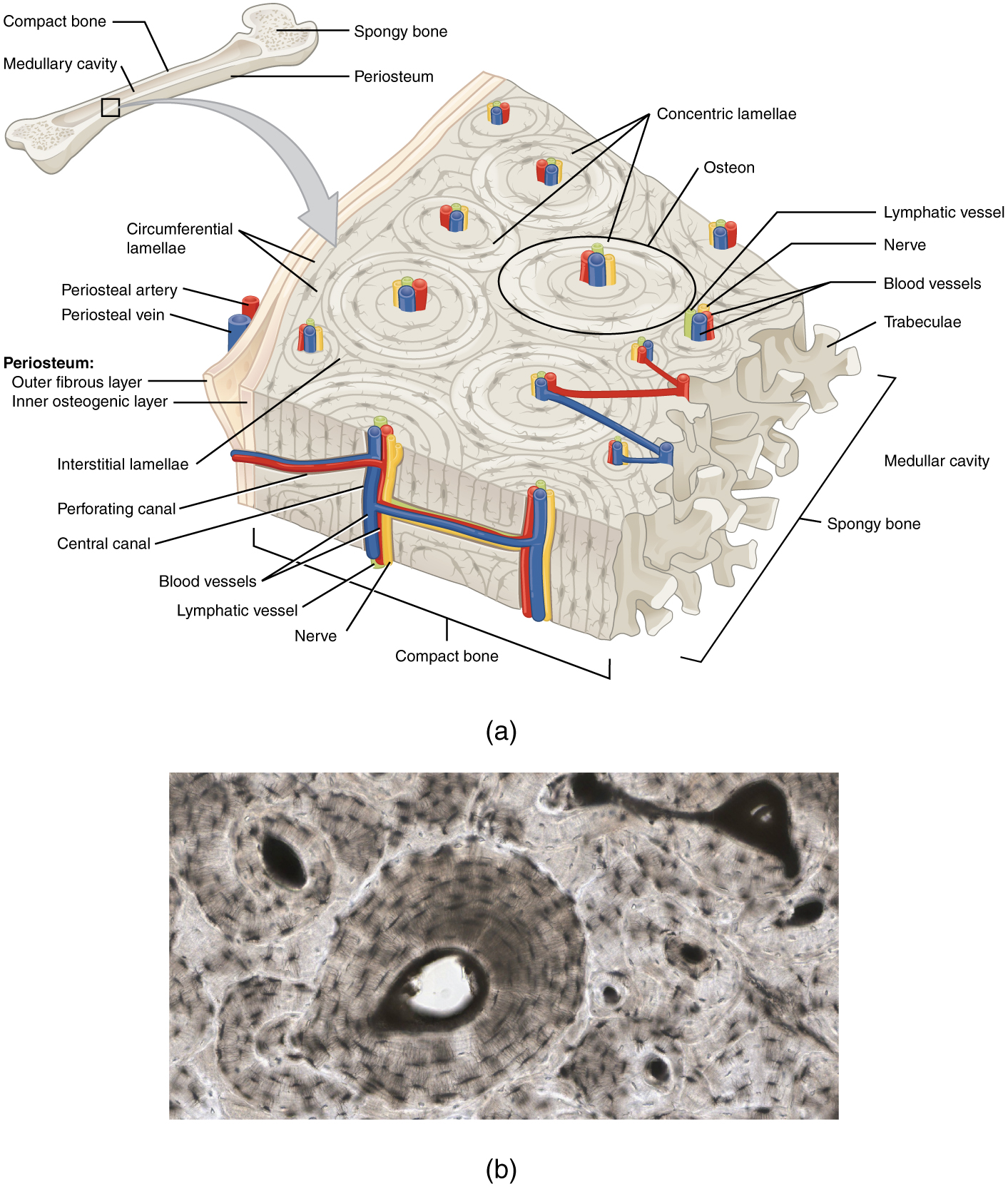
A generic long bone is shown at the top of this illustration. The bone is split in half lengthwise to show its internal anatomy. The outer gray covering of the bone is labeled the periosteum. Within the periosteum is a thin layer of compact bone. The compact bone surrounds a central cavity called the medullary cavity. The medullary cavity is filled with spongy bone at the two epiphyses. A callout box shows that the main image is zooming in on the compact bone on the left side of the bone. On the main image, the periosteum is being peeled back to show its two layers. The outer layer of the periosteum is the outer fibrous layer. This layer has a periosteal artery and a periosteal vein running along its outside edge. The inner layer of the periosteum is labeled the inner osteogenic layer. The compact bone lies to the right of the periosteum and occupies the majority of the main image. Two flat layers of compact bone line the inner surface of the ostegenic periosteum. These sheets of compact bone are called the circumferential lamellae. The majority of the compact bone has lamellae running perpendicular to that of the circumferential lamellae. These concentric lamellae are arranged in a series of concentric tubes. There are small cavities between the layers of concentric lamellae called lacunae. The centermost concentric lamella surrounds a hollow central canal. A blue vein, a red artery, a yellow nerve and a green lymph vessel run vertically through the central canal. A set of concentric lamellae, its associated lacunae and the vessels and nerves of the central canal are collectively called an osteon. The front edge of the diagram shows a longitudinal cross section of one of the osteons. The vessels and nerve are visible running through the center of the osteon throughout its length. In addition, blood vessels can run from the periosteum through the sides of the osteons and connect with the vessels of the central canal. The blood vessels travel through the sides of the osteons via a perforating canal. The open areas between neighboring osteons are also filled with compact bone. This “filler” bone is referred to as the interstitial lamellae. At the far right of the compact bone, the edge of the spongy bone is visible. The spongy bone is a series of crisscrossing bony arches called trabeculae. There are many open spaces between the trabeculae, giving the spongy bone its sponge-like appearance.
Image: OpenStax / University of Hawaiʻi adapted teaching figures; original contributors credited at source · Source / original · CC BY 4.0 — figure credits at sourceNumbered markers and focused views are Mediveerse learning overlays; the original reference is linked above.
Compact bone: osteon
Understand what you see.
Provides rigid load-bearing support while maintaining living cells through vascular canal systems.
Zoom into the original labels. Practise pointing out each structure, then explain it aloud. Diagrams are teaching references; specimen appearance and slide staining vary.
How to recognise it.
- Concentric lamellae around central canals
- Osteocyte lacunae between lamellae
- Canaliculi connect lacunar spaces
Point them out.
Tap a number to open its reference, zoom in and locate the structure. Numbers stay the same across views. This key follows landmarks visible in the reference images.
Think first. Then reveal.
Say your answer aloud before opening the suggested response.
01How do you identify compact bone: osteon?+
Concentric lamellae around central canals. Osteocyte lacunae between lamellae. Canaliculi connect lacunar spaces.
02Differentiate Haversian and Volkmann canals.+
Haversian canals run longitudinally within osteons. Volkmann canals connect vascular channels across osteons and are not surrounded by the same concentric lamellar pattern.
03Where are osteocytes found?+
Within lacunae, with processes extending through canaliculi.
04Which structures should you point out on this reference?+
Central canal; Concentric lamellae; Lacunae and canaliculi. Provides rigid load-bearing support while maintaining living cells through vascular canal systems.
Read the references.
Credited anatomical plates, microscopy and university teaching references. Individual image credits appear below the viewer. Historical plates can use older terminology; use the lesson text for current names.