 MEDIVEERSETHE PRACTICAL HUB
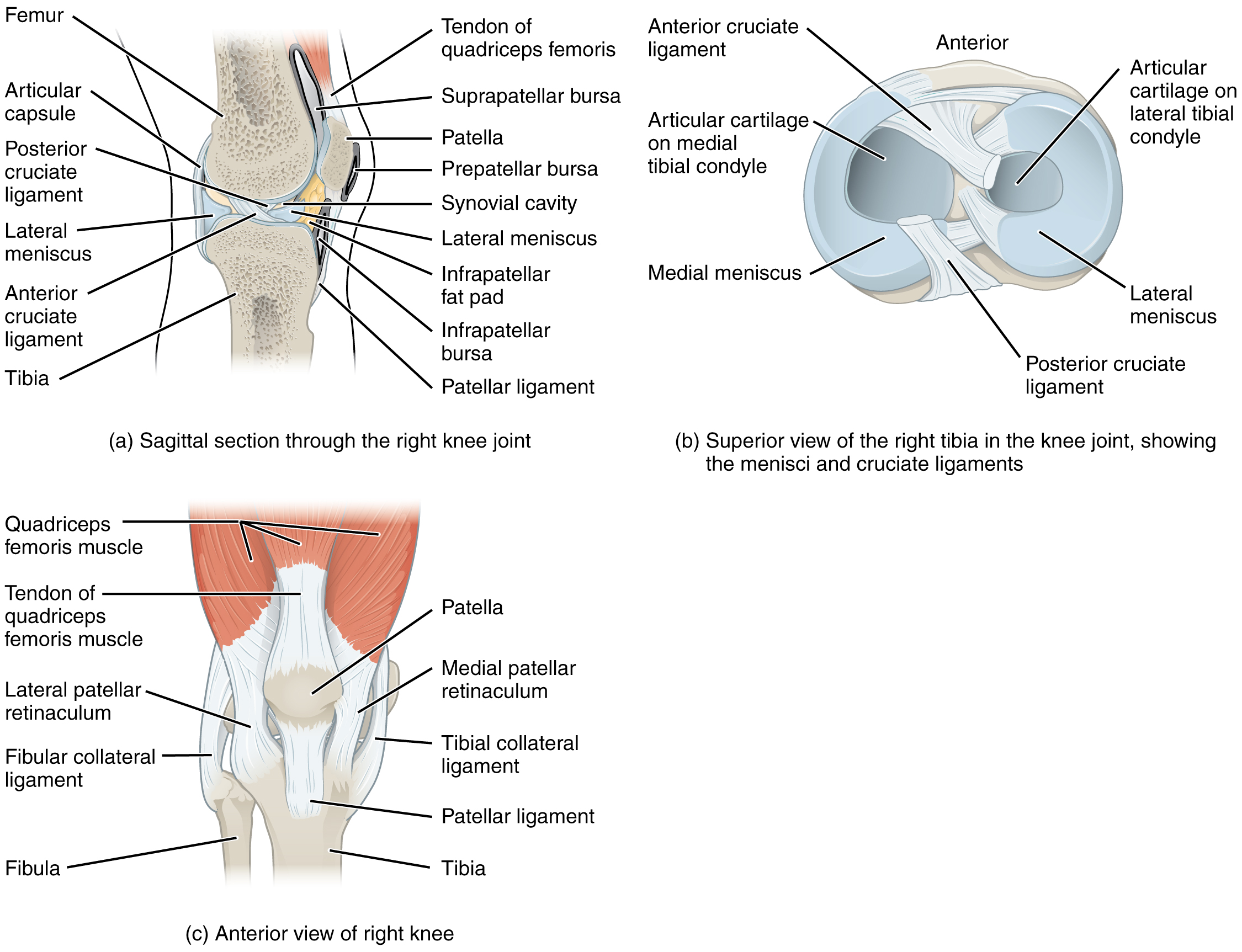
Locate medial meniscus on this anatomical reference. Read the original plate labels to confirm the view and adjacent structures.
Zoom in, then drag to inspect. Keyboard: + / − to zoom, arrows to pan, 0 to reset. Tap the numbered markers to explore parts.
This image shows the different views of the knee joint. The top, left panel shows the sagittal view of the right knee joint. The top, left panel shows the superior view of the right tibia, identifying the ligaments. The bottom, right panel shows the anterior view of the right knee.
Image: OpenStax / University of Hawaiʻi adapted teaching figures; original contributors credited at source · Source / original · CC BY 4.0 — figure credits at sourceNumbered markers and focused views are Mediveerse learning overlays; the original reference is linked above.
Knee joint
Understand what you see.
Modified hinge joint permitting flexion, extension and rotation depending on position.
Zoom into the original labels. Practise pointing out each structure, then explain it aloud. Diagrams are teaching references; specimen appearance and slide staining vary.
How to recognise it.
- Femoral condyles meet tibial plateaux
- Menisci deepen contact
- Patella articulates with femur
Point them out.
Tap a number to open its reference, zoom in and locate the structure. Numbers stay the same across views. This key follows landmarks visible in the reference images.
Think first. Then reveal.
Say your answer aloud before opening the suggested response.
01How do you identify knee joint?+
Femoral condyles meet tibial plateaux. Menisci deepen contact. Patella articulates with femur.
02How do cruciates restrain movement?+
ACL mainly limits anterior tibial translation relative to femur; PCL mainly limits posterior tibial translation.
03Why is the medial meniscus relatively less mobile?+
It has stronger capsular attachments, including attachment to the deep medial collateral region.
04What is its main functional or developmental significance?+
Modified hinge joint permitting flexion, extension and rotation depending on position. Key structures to recognise: Menisci; Cruciate ligaments; Collateral ligaments.
Read the references.
Credited anatomical plates, microscopy and university teaching references. Individual image credits appear below the viewer. Historical plates can use older terminology; use the lesson text for current names.