 MEDIVEERSETHE PRACTICAL HUB
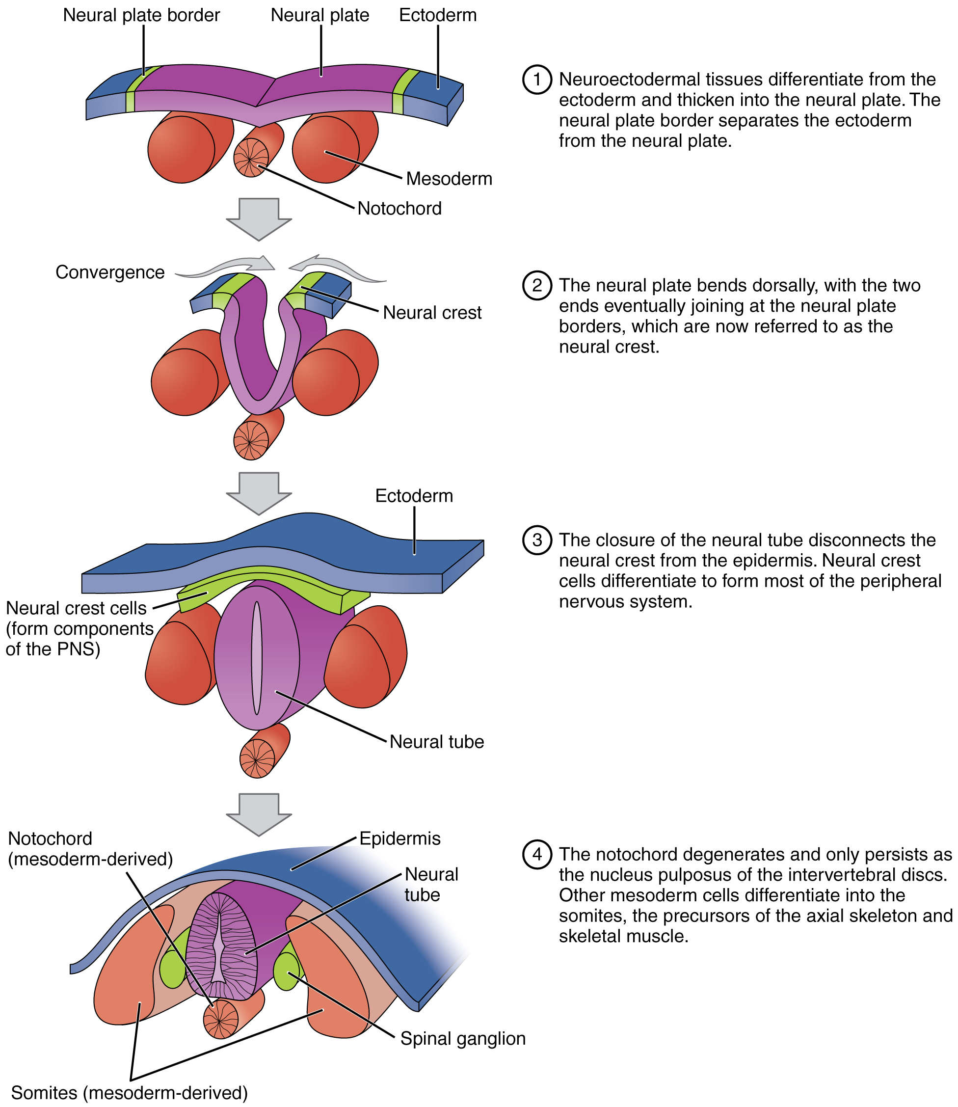
Locate neural folds on this anatomical reference. Read the original plate labels to confirm the view and adjacent structures.
Zoom in, then drag to inspect. Keyboard: + / − to zoom, arrows to pan, 0 to reset. Tap the numbered markers to explore parts.
neurulation: labelled anatomical figure. Original source colours and labels retained.
Image: OpenStax / University of Hawaiʻi adapted teaching figures; original contributors credited at source · Source / original · CC BY 4.0 — figure credits at sourceNumbered markers and focused views are Mediveerse learning overlays; the original reference is linked above.
Neural-tube defects: developmental basis
Understand what you see.
Relates neurulation failure to congenital abnormalities; the illustration shows normal closure for comparison.
Zoom into the original labels. Practise pointing out each structure, then explain it aloud. Diagrams are teaching references; specimen appearance and slide staining vary.
How to recognise it.
- Abnormal neural-tube closure spectrum
- Cranial and spinal forms differ
- Severity depends on location and tissue involvement
Point them out.
Tap a number to open its reference, zoom in and locate the structure. Numbers stay the same across views. This key follows landmarks visible in the reference images.
Think first. Then reveal.
Say your answer aloud before opening the suggested response.
01How do you identify neural-tube defects: developmental basis?+
Abnormal neural-tube closure spectrum. Cranial and spinal forms differ. Severity depends on location and tissue involvement.
02Is spina bifida always an open neural lesion?+
No. The term covers a spectrum, including vertebral-arch defects with or without exposed neural tissue.
03Why compare with a normal neurulation diagram?+
Understanding plate elevation, fold fusion and surface separation makes the developmental failure easier to explain without assuming all defects have the same mechanism.
04Which structures should you point out on this reference?+
Cranial neuropore; Caudal neuropore; Neural folds. Relates neurulation failure to congenital abnormalities; the illustration shows normal closure for comparison.
Read the references.
Credited anatomical plates, microscopy and university teaching references. Individual image credits appear below the viewer. Historical plates can use older terminology; use the lesson text for current names.