 MEDIVEERSETHE PRACTICAL HUB
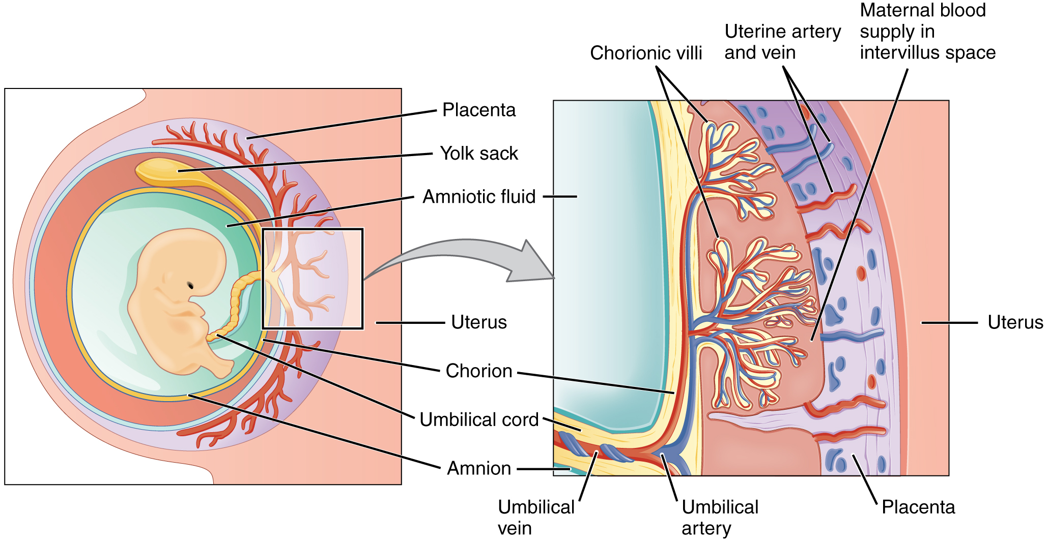
Locate chorionic villi on this anatomical reference. Read the original plate labels to confirm the view and adjacent structures.
Zoom in, then drag to inspect. Keyboard: + / − to zoom, arrows to pan, 0 to reset. Tap the numbered markers to explore parts.
placenta: labelled anatomical figure. Original source colours and labels retained.
Image: OpenStax / University of Hawaiʻi adapted teaching figures; original contributors credited at source · Source / original · CC BY 4.0 — figure credits at sourceNumbered markers and focused views are Mediveerse learning overlays; the original reference is linked above.
Placental exchange barrier
Understand what you see.
Permits regulated maternal–fetal exchange without a normal direct mixing of the two circulations.
Zoom into the original labels. Practise pointing out each structure, then explain it aloud. Diagrams are teaching references; specimen appearance and slide staining vary.
How to recognise it.
- Maternal blood occupies intervillous spaces
- Fetal blood remains in villous capillaries
- Barrier composition changes with gestation
Point them out.
Tap a number to open its reference, zoom in and locate the structure. Numbers stay the same across views. This key follows landmarks visible in the reference images.
Think first. Then reveal.
Say your answer aloud before opening the suggested response.
01How do you identify placental exchange barrier?+
Maternal blood occupies intervillous spaces. Fetal blood remains in villous capillaries. Barrier composition changes with gestation.
02Does maternal blood normally enter umbilical vessels directly?+
No. Maternal and fetal blood are normally separated by the placental interface.
03Is the barrier absolutely impermeable?+
No. Many substances, antibodies and some pathogens can cross; permeability is selective and changes with development.
04Which structures should you point out on this reference?+
Syncytiotrophoblast; Villous stroma; Fetal capillary endothelium. Permits regulated maternal–fetal exchange without a normal direct mixing of the two circulations.
Read the references.
Credited anatomical plates, microscopy and university teaching references. Individual image credits appear below the viewer. Historical plates can use older terminology; use the lesson text for current names.