 MEDIVEERSETHE PRACTICAL HUB
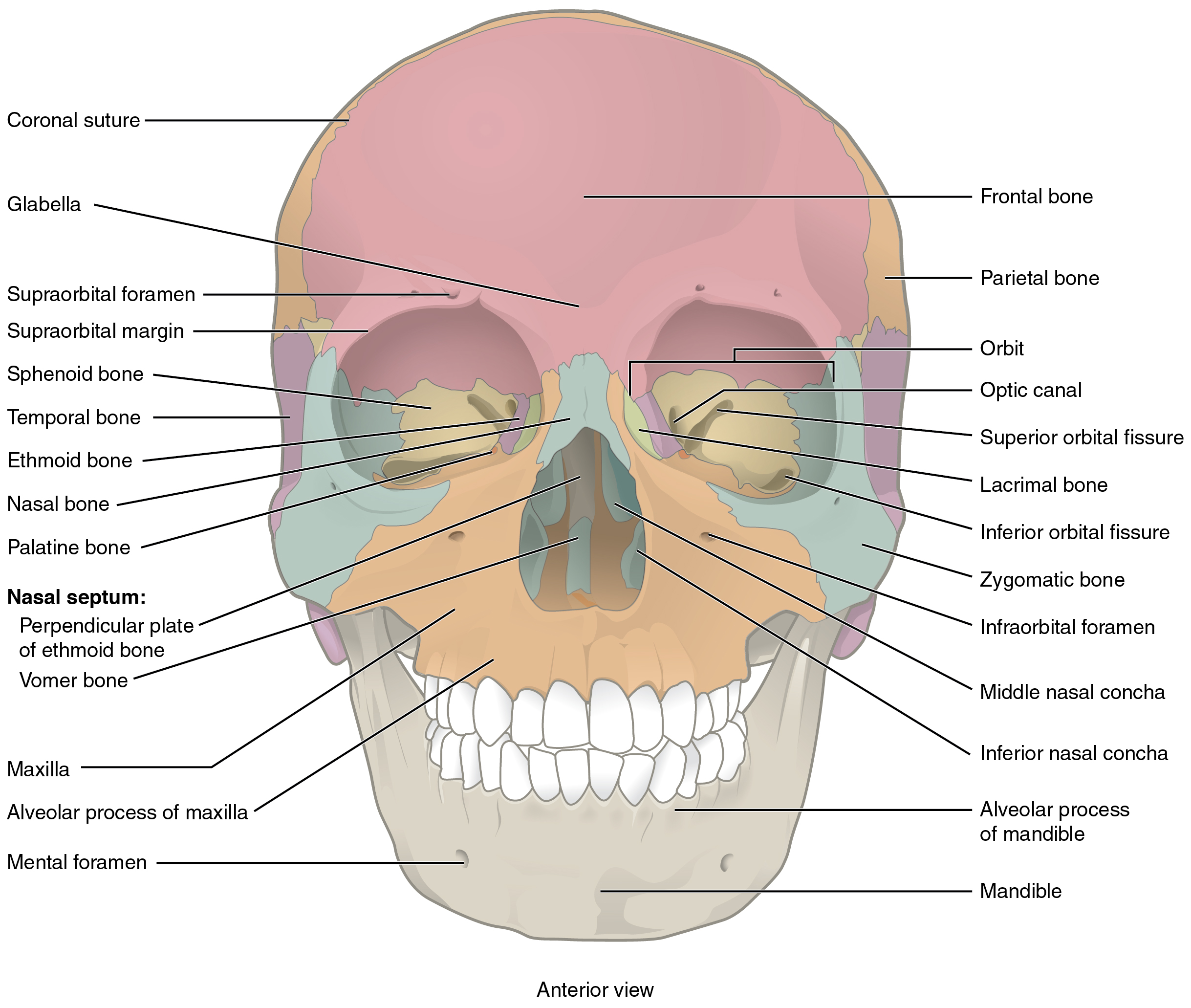
Forms the forehead and roof of the orbit. Contains frontal air sinuses in the adult. Meets the parietal bones at the coronal suture.
Study this structure →Zoom in, then drag to inspect. Keyboard: + / − to zoom, arrows to pan, 0 to reset. Tap the numbered markers to explore parts.
Skull: views & sutures: labelled teaching diagram. Original labels and colours retained.
Image: OpenStax College / university open textbook · Source / original · CC BY 4.0Numbered markers and focused views are Mediveerse learning overlays; the original reference is linked above.
Skull: views & sutures
Understand what you see.
Protects the brain and supports facial and sensory structures.
Zoom into the original labels. Practise pointing out each structure, then explain it aloud. Diagrams are teaching references; specimen appearance and slide staining vary.
How to recognise it.
- Cranial bones surround the brain; facial bones form the face
- Coronal suture separates frontal and parietal bones
- Sagittal suture lies between the parietal bones
Point them out.
Tap a number to open its reference, zoom in and locate the structure. Numbers stay the same across views. This key follows landmarks visible in the reference images.
Think first. Then reveal.
Say your answer aloud before opening the suggested response.
Examiner-style prompts: identify the structure, explain its relations, then answer the applied follow-up. These are revision questions, not authenticated past papers.
01How would you identify skull: views & sutures?+
Cranial bones surround the brain; facial bones form the face. Coronal suture separates frontal and parietal bones. Sagittal suture lies between the parietal bones.
02Where do the coronal and sagittal sutures meet?+
At bregma, corresponding to the site of the anterior fontanelle in the infant skull.
03How many bones form the adult skull, excluding auditory ossicles and hyoid?+
Twenty-two: eight cranial and fourteen facial bones.
04Which bones form the anterior fontanelle boundaries?+
The two frontal and two parietal bones at their meeting region in infancy.
05Differentiate bregma from lambda.+
Bregma is the coronal–sagittal junction; lambda is the sagittal–lambdoid junction.
06Why should you identify the viewing plane before a skull spotter?+
A landmark may be visible in only one view; state anterior, lateral, inferior or endocranial orientation before naming it.
Read the references.
Credited anatomical plates, microscopy and university teaching references. Individual image credits appear below the viewer. Historical plates can use older terminology; use the lesson text for current names.