 MEDIVEERSETHE PRACTICAL HUB
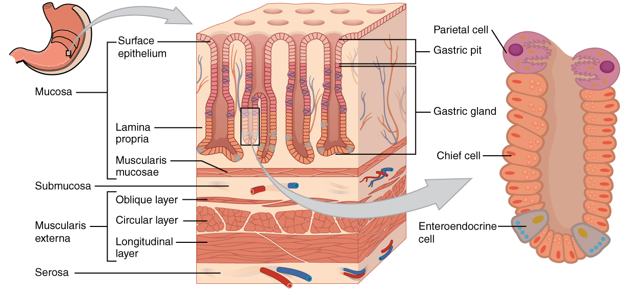
Mucosal invagination leading to gastric glands.
Zoom in, then drag to inspect. Keyboard: + / − to zoom, arrows to pan, 0 to reset. Tap the numbered markers to explore parts.
stomach: labelled anatomical figure. Original source colours and labels retained.
Image: OpenStax / University of Hawaiʻi adapted teaching figures; original contributors credited at source · Source / original · CC BY 4.0 — figure credits at sourceNumbered markers and focused views are Mediveerse learning overlays; the original reference is linked above.
Stomach: gastric mucosa
Understand what you see.
Produces protective mucus, acid, intrinsic factor and digestive precursors according to region.
Zoom into the original labels. Practise pointing out each structure, then explain it aloud. Diagrams are teaching references; specimen appearance and slide staining vary.
How to recognise it.
- Gastric pits open into glands
- Simple columnar surface mucous cells
- No villi
Point them out.
Tap a number to open its reference, zoom in and locate the structure. Numbers stay the same across views. This key follows landmarks visible in the reference images.
Think first. Then reveal.
Say your answer aloud before opening the suggested response.
01How do you identify stomach: gastric mucosa?+
Gastric pits open into glands. Simple columnar surface mucous cells. No villi.
02What do parietal cells secrete?+
Hydrochloric acid and intrinsic factor.
03What do chief cells secrete?+
Pepsinogen and gastric lipase; cell distribution differs by gastric region.
04Which structures should you point out on this reference?+
Gastric pits; Parietal cells; Chief cells. Produces protective mucus, acid, intrinsic factor and digestive precursors according to region.
Read the references.
Credited anatomical plates, microscopy and university teaching references. Individual image credits appear below the viewer. Historical plates can use older terminology; use the lesson text for current names.