 MEDIVEERSETHE PRACTICAL HUB
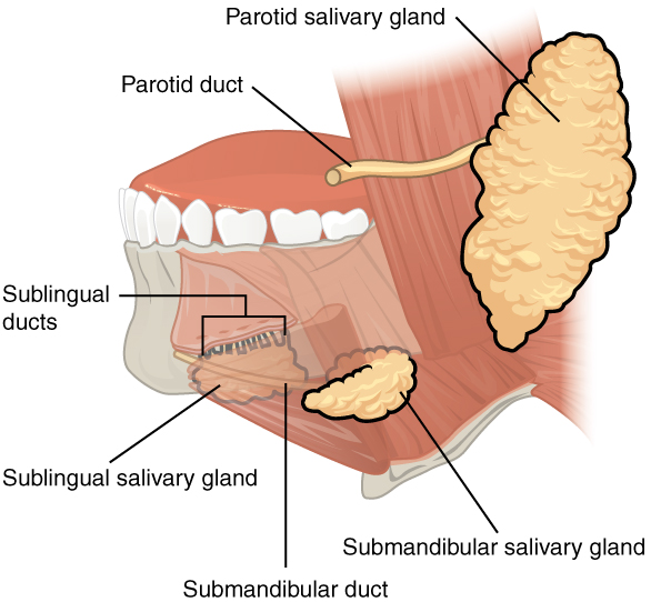
Locate submandibular gland on this anatomical reference. Read the original plate labels to confirm the view and adjacent structures.
Zoom in, then drag to inspect. Keyboard: + / − to zoom, arrows to pan, 0 to reset. Tap the numbered markers to explore parts.
salivary: labelled anatomical figure. Original source colours and labels retained.
Image: OpenStax / University of Hawaiʻi adapted teaching figures; original contributors credited at source · Source / original · CC BY 4.0 — figure credits at sourceNumbered markers and focused views are Mediveerse learning overlays; the original reference is linked above.
Submandibular gland
Understand what you see.
Produces mixed, predominantly serous saliva.
Zoom into the original labels. Practise pointing out each structure, then explain it aloud. Diagrams are teaching references; specimen appearance and slide staining vary.
How to recognise it.
- Mixed gland mainly below mandible
- Superficial and deep parts wrap around posterior mylohyoid border
- Duct runs forward in mouth floor
Point them out.
Tap a number to open its reference, zoom in and locate the structure. Numbers stay the same across views. This key follows landmarks visible in the reference images.
Think first. Then reveal.
Say your answer aloud before opening the suggested response.
Examiner-style prompts: identify the structure, explain its relations, then answer the applied follow-up. These are revision questions, not authenticated past papers.
01Identify submandibular gland and give its main role.+
Mixed gland mainly below mandible. Superficial and deep parts wrap around posterior mylohyoid border. Duct runs forward in mouth floor. Produces mixed, predominantly serous saliva.
02Which structure separates its superficial and deep parts?+
Mylohyoid; the parts remain continuous around its posterior free border.
03Trace parasympathetic supply.+
VII chorda tympani fibres join the lingual nerve, synapse in submandibular ganglion, and postganglionic fibres reach the gland.
04Point out three important relations and explain their significance.+
Superficial part; Deep part; Mylohyoid posterior border. Produces mixed, predominantly serous saliva. Use the reference plate to locate the region; the relationship map organises the associations and is not drawn to scale.
Read the references.
Credited anatomical plates, microscopy and university teaching references. Individual image credits appear below the viewer. Historical plates can use older terminology; use the lesson text for current names.