 MEDIVEERSETHE PRACTICAL HUB
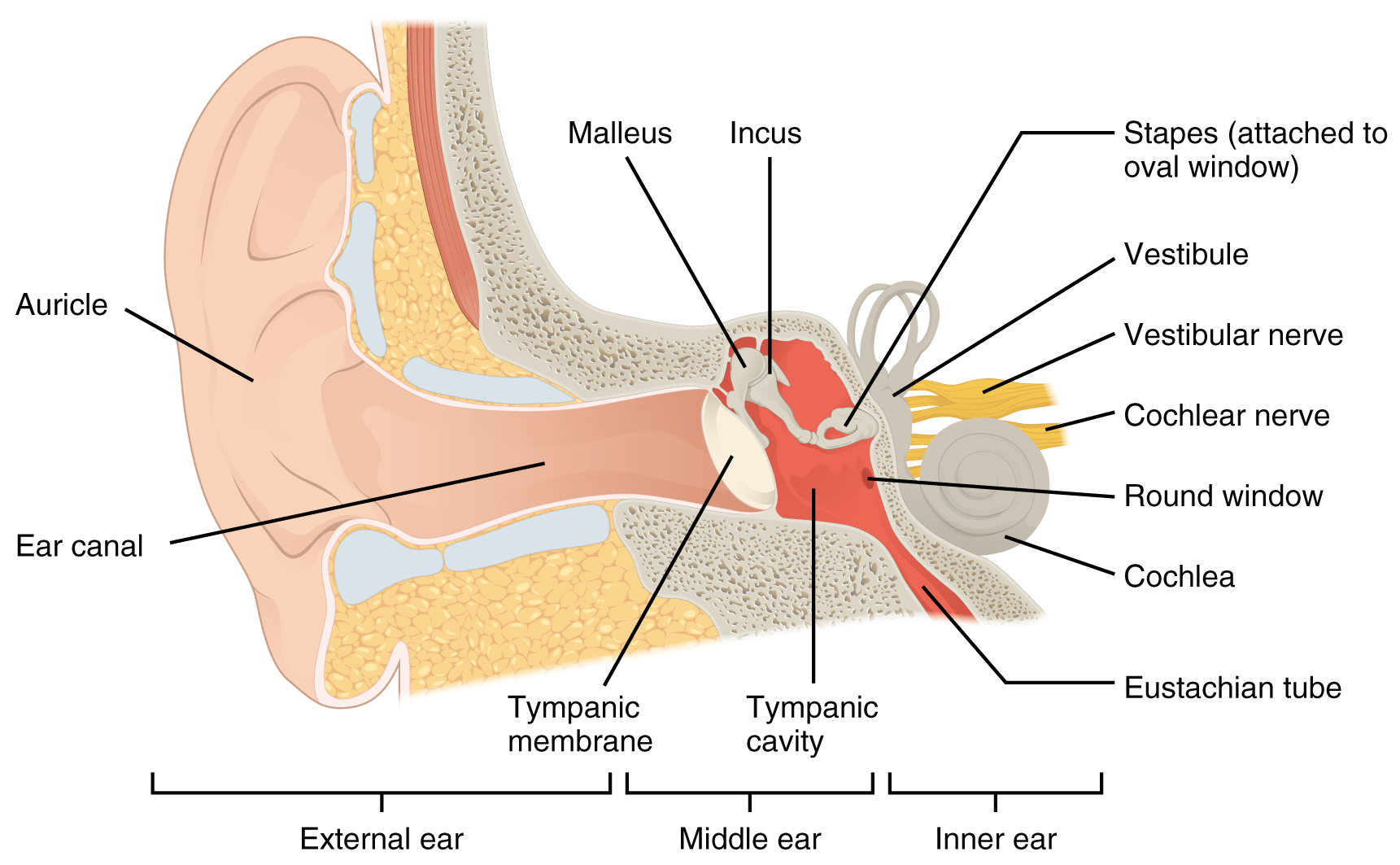
Air-filled temporal-bone cavity. Contains auditory ossicles. Communicates with nasopharynx and mastoid system.
Zoom in, then drag to inspect. Keyboard: + / − to zoom, arrows to pan, 0 to reset. Tap the numbered markers to explore parts.
Tympanic cavity: magnified reference view with a numbered learning marker. The complete original plate is available in the next view.
Image: OpenStax College · Source / original · CC BY 4.0Numbered markers and focused views are Mediveerse learning overlays; the original reference is linked above.
Tympanic cavity
Understand what you see.
Conducts sound through the middle-ear ossicles.
Zoom into the original labels. Practise pointing out each structure, then explain it aloud. Diagrams are teaching references; specimen appearance and slide staining vary.
How to recognise it.
- Air-filled temporal-bone cavity
- Contains auditory ossicles
- Communicates with nasopharynx and mastoid system
Point them out.
Tap a number to open its reference, zoom in and locate the structure. Numbers stay the same across views. This key follows landmarks visible in the reference images.
Think first. Then reveal.
Say your answer aloud before opening the suggested response.
Examiner-style prompts: identify the structure, explain its relations, then answer the applied follow-up. These are revision questions, not authenticated past papers.
01Identify tympanic cavity and give its main role.+
Air-filled temporal-bone cavity. Contains auditory ossicles. Communicates with nasopharynx and mastoid system. Conducts sound through the middle-ear ossicles.
02Name clinically important wall relations.+
Roof relates to middle cranial fossa; floor to jugular bulb; anterior wall to carotid canal and auditory tube; medial wall to labyrinth and facial canal.
03Which nerve crosses the cavity without supplying hearing?+
Chorda tympani, carrying taste and parasympathetic fibres, crosses the middle ear.
04Point out three important relations and explain their significance.+
Roof: tegmen tympani; Medial: labyrinthine wall; Posterior: aditus to mastoid antrum. Conducts sound through the middle-ear ossicles. Use the reference plate to locate the region; the relationship map organises the associations and is not drawn to scale.
Read the references.
Credited anatomical plates, microscopy and university teaching references. Individual image credits appear below the viewer. Historical plates can use older terminology; use the lesson text for current names.