 MEDIVEERSETHE PRACTICAL HUB
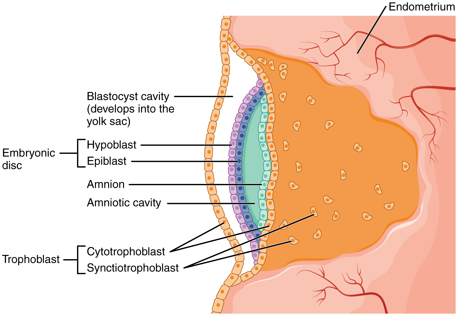
Epiblast gives rise to all three definitive germ layers.
Zoom in, then drag to inspect. Keyboard: + / − to zoom, arrows to pan, 0 to reset. Tap the numbered markers to explore parts.
disc: labelled anatomical figure. Original source colours and labels retained.
Image: OpenStax / University of Hawaiʻi adapted teaching figures; original contributors credited at source · Source / original · CC BY 4.0 — figure credits at sourceNumbered markers and focused views are Mediveerse learning overlays; the original reference is linked above.
Yolk sac
Understand what you see.
Contributes to early nutrient transfer, early haematopoiesis and germ-cell-related developmental pathways.
Zoom into the original labels. Practise pointing out each structure, then explain it aloud. Diagrams are teaching references; specimen appearance and slide staining vary.
How to recognise it.
- Extraembryonic sac ventral to early disc
- Communicates transiently with midgut
- Related to early blood formation
Point them out.
Tap a number to open its reference, zoom in and locate the structure. Numbers stay the same across views. This key follows landmarks visible in the reference images.
Think first. Then reveal.
Say your answer aloud before opening the suggested response.
01How do you identify yolk sac?+
Extraembryonic sac ventral to early disc. Communicates transiently with midgut. Related to early blood formation.
02Does the human yolk sac contain a large yolk store?+
No. Its name reflects comparative embryology; human development relies on other nutritional routes.
03What connects it with the midgut?+
Vitelline duct, which normally regresses; persistence can produce anomalies such as a Meckel diverticulum.
04Which structures should you point out on this reference?+
Yolk-sac lining; Vitelline connection; Extraembryonic mesoderm. Contributes to early nutrient transfer, early haematopoiesis and germ-cell-related developmental pathways.
Read the references.
Credited anatomical plates, microscopy and university teaching references. Individual image credits appear below the viewer. Historical plates can use older terminology; use the lesson text for current names.