Verify
Confirm identity, date, orientation and image quality.
 MEDIVEERSETHE PRACTICAL HUB
OBSERVE / REASON / EXPLAIN
Orient a panoramic teaching radiograph and explain its limitations.
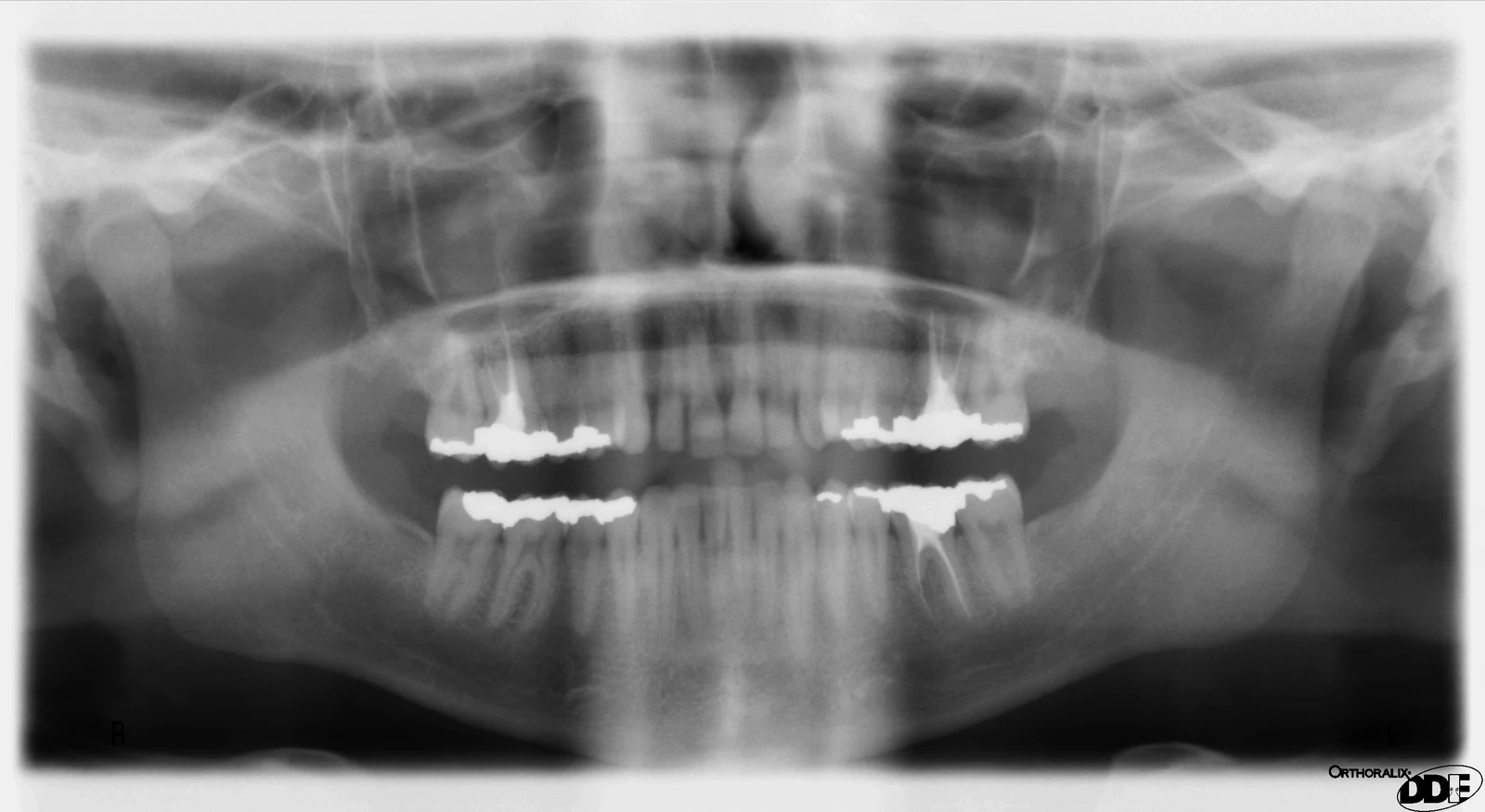
The upper anterior dentition in the panoramic projection. This is a reference for orientation, not a diagnosis.
Zoom in, then drag to inspect. Keyboard: + / − to zoom, arrows to pan, 0 to reset. Tap the numbered markers to explore parts.
panoramic radiograph — reference view
Image: Author credited at source · Source / original · Public domainResized and converted to WebP; original colours retained.
Panoramic imaging gives a two-dimensional overview with magnification, superimposition and positioning effects.
Confirm identity, date, orientation and image quality.
Follow a systematic anatomical search pattern.
Select additional imaging only when justified by the clinical question.
Try answering aloud, then open the suggested answer. Examiner questions vary.
No. It is a two-dimensional projection.
Yes. Positioning can cause distortion or obscure structures.
No. Correlate with history and examination.
Selection should be based on individual clinical need and the appropriate guidance.