Red-cell assessment
Describe size, colour and shape without assigning a diagnosis from one cell.
 MEDIVEERSETHE PRACTICAL HUB
OBSERVE / REASON / EXPLAIN
Recognise the formed elements and describe a blood film systematically.
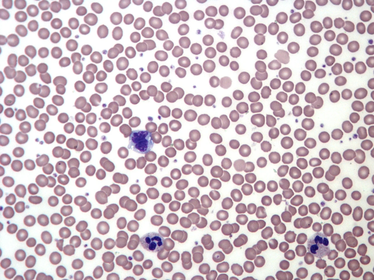
An anucleate red cell with central pallor in this teaching film.
Zoom in, then drag to inspect. Keyboard: + / − to zoom, arrows to pan, 0 to reset. Tap the numbered markers to explore parts.
blood smear — reference view
Image: Keith Chambers · Source / original · CC BY-SA 3.0Resized and converted to WebP; original colours retained.
A stained thin blood film separates cells for microscopic assessment; morphology must be interpreted with counts and clinical context.
Describe size, colour and shape without assigning a diagnosis from one cell.
Assess lineage features and distribution; a representative area is essential.
Review the CBC, sample quality and clinical history. The reference is a normal adult film.
Try answering aloud, then open the suggested answer. Examiner questions vary.
No. Counts, history and additional tests are often required.
Variation in red-cell size.
Variation in red-cell shape.
Thick areas, poor staining and artefacts can distort apparent morphology.