Recognise
Identify the viewing head and illumination system.
 MEDIVEERSETHE PRACTICAL HUB
LOOK CLOSELY / UNDERSTAND CLEARLY
Recognise a dermatoscope and explain its role in supervised skin assessment.
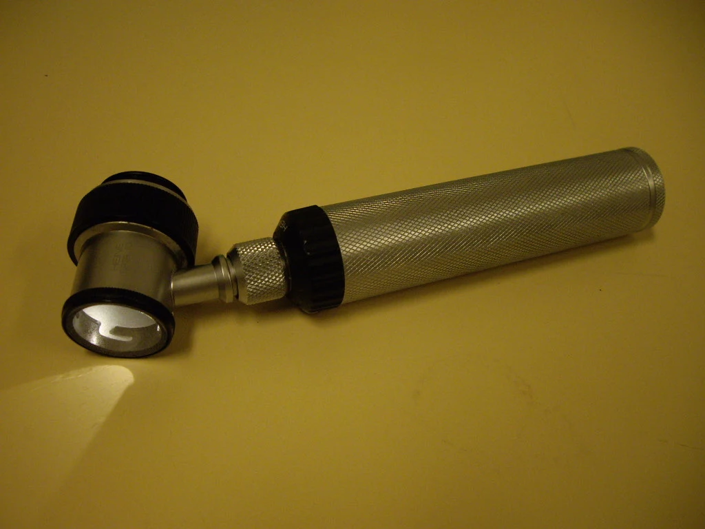
The magnified viewing and illumination head of this reference dermatoscope.
Zoom in, then drag to inspect. Keyboard: + / − to zoom, arrows to pan, 0 to reset. Tap the numbered markers to explore parts.
dermatoscope — reference view
Image: Frank33 · Source / original · CC BY-SA 3.0Resized and converted to WebP; original colours retained.
Magnification with controlled illumination helps examine surface and subsurface skin structures.
Identify the viewing head and illumination system.
Record lesion site, distribution and morphology before dermoscopy.
Dermoscopy supplements history and examination; suspicious findings require appropriate specialist assessment.
Try answering aloud, then open the suggested answer. Examiner questions vary.
No. Histology examines sampled tissue microscopically.
No. Pattern and clinical context matter.
The arrangement and sites of lesions can narrow the differential.
No. It cannot provide all clinical information.