Identify
Choose the appropriate speculum and recognise the viewing optics.
 MEDIVEERSETHE PRACTICAL HUB
LOOK CLOSELY / UNDERSTAND CLEARLY
Identify an otoscope and explain what a supervised ear examination assesses.
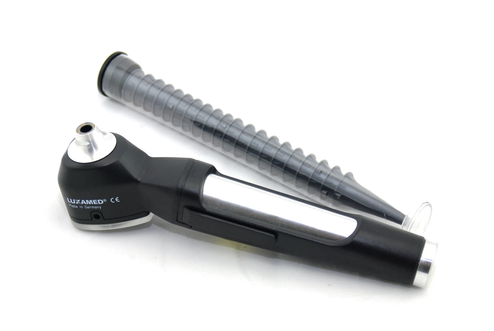
The examination head’s cone attachment. A separate ear speculum is shown above.
Zoom in, then drag to inspect. Keyboard: + / − to zoom, arrows to pan, 0 to reset. Tap the numbered markers to explore parts.
otoscope — reference view
Image: GO (MedPlus Medizintechnik GmbH) · Source / original · CC BY 4.0Resized and converted to WebP; original colours retained.
Illumination and magnification allow inspection of the external canal and tympanic membrane when visible.
Choose the appropriate speculum and recognise the viewing optics.
Describe the canal and visible tympanic membrane without forcing an obstructed view.
Correlate with symptoms and hearing assessment.
Try answering aloud, then open the suggested answer. Examiner questions vary.
No. The tympanic membrane limits the view.
Fit affects comfort and the field of view.
No. Hearing loss has several conductive and sensorineural causes.
No. Stop and seek appropriate clinical assessment.