Describe
Report colour, shape and arrangement before interpretation.
 MEDIVEERSETHE PRACTICAL HUB
OBSERVE / REASON / EXPLAIN
Describe staining reaction, morphology and arrangement on a validated teaching image.
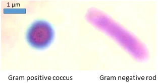
The rounded purple-staining organism in this reference.
Zoom in, then drag to inspect. Keyboard: + / − to zoom, arrows to pan, 0 to reset. Tap the numbered markers to explore parts.
gram stain — reference view
Image: Mikael Häggström, M.D. Author info - Reusing images- Conflicts of interest: None Mikael Häggström, M.D. · Source / original · CC0Resized and converted to WebP; original colours retained.
Differential staining reflects cell-envelope behaviour under the method; appearance does not determine species alone.
Report colour, shape and arrangement before interpretation.
Mixed flora, ageing cultures, technique and sample quality can complicate results.
Use the sample site, culture and other validated tests.
Try answering aloud, then open the suggested answer. Examiner questions vary.
No. It is one preliminary characteristic.
It changes retention of the staining complexes.
Yes. Mixed specimens need careful interpretation.
No. Follow the laboratory’s risk assessment and biosafety protocol.