Identify
Observe the probe shape and orientation marker where visible.
 MEDIVEERSETHE PRACTICAL HUB
LOOK CLOSELY / UNDERSTAND CLEARLY
Recognise an ultrasound probe and explain image generation and probe selection.
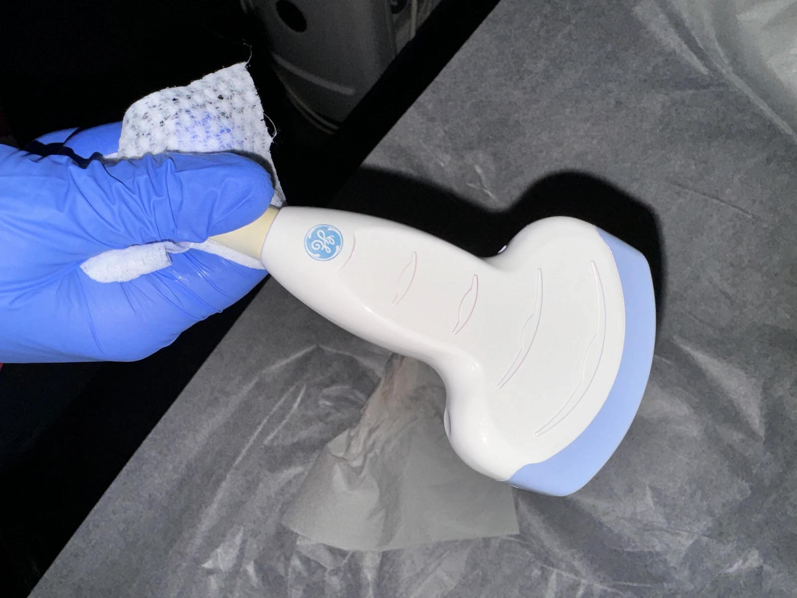
The patient-contact acoustic face of the reference probe; probe geometry varies by design.
Zoom in, then drag to inspect. Keyboard: + / − to zoom, arrows to pan, 0 to reset. Tap the numbered markers to explore parts.
ultrasound probe — reference view
Image: Harrison Keely · Source / original · CC BY 4.0Resized and converted to WebP; original colours retained.
A transducer sends and receives acoustic signals; processed echoes generate an image.
Observe the probe shape and orientation marker where visible.
Frequency, penetration and resolution are related.
Use clinically appropriate examinations and validated cleaning procedures.
Try answering aloud, then open the suggested answer. Examiner questions vary.
No. It uses acoustic energy.
No. Geometry and frequency range suit different applications.
It links the displayed image to the patient’s anatomy.
No. Ultrasound should still be used appropriately and with prudent exposure.