 MEDIVEERSETHE PRACTICAL HUB
See the instrument.
Focus on the detail.
From the trial frame to the slit lamp. Learn what each instrument does, identify its parts and practise explaining the science behind it.
Back to Practical Hub24 instruments to explore
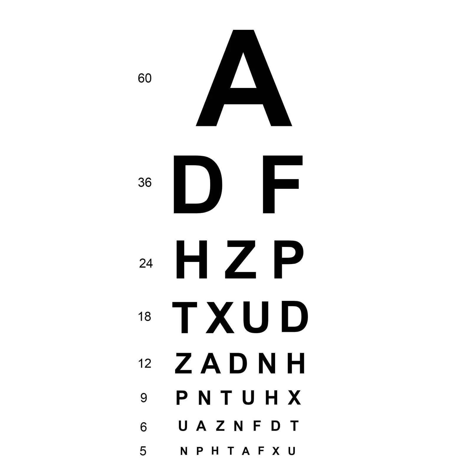
Snellen visual-acuity chart
Assesses distance visual acuity under standardised viewing conditions.
Identify · Locate · Practise 4 viva questions · Zoomable reference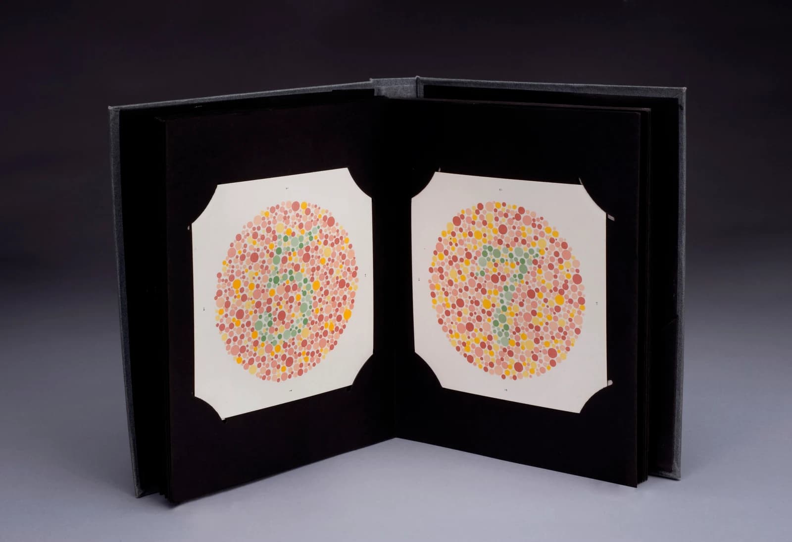
Ishihara colour-vision plates
Screens principally for red–green colour-vision deficiency.
Identify · Locate · Practise 4 viva questions · Zoomable reference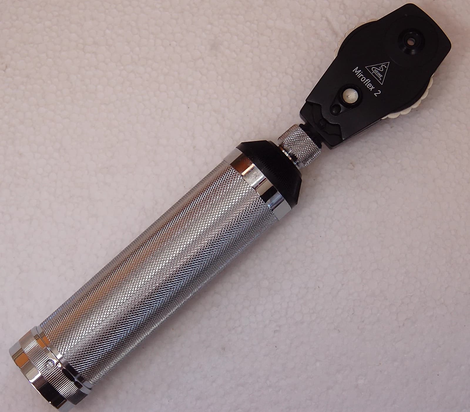
Direct ophthalmoscope
Examines the ocular fundus, including the optic disc and retinal vessels.
Identify · Locate · Practise 4 viva questions · Zoomable reference
Visual-field perimeter
Maps the extent and sensitivity of the visual field.
Identify · Locate · Practise 4 viva questions · Zoomable reference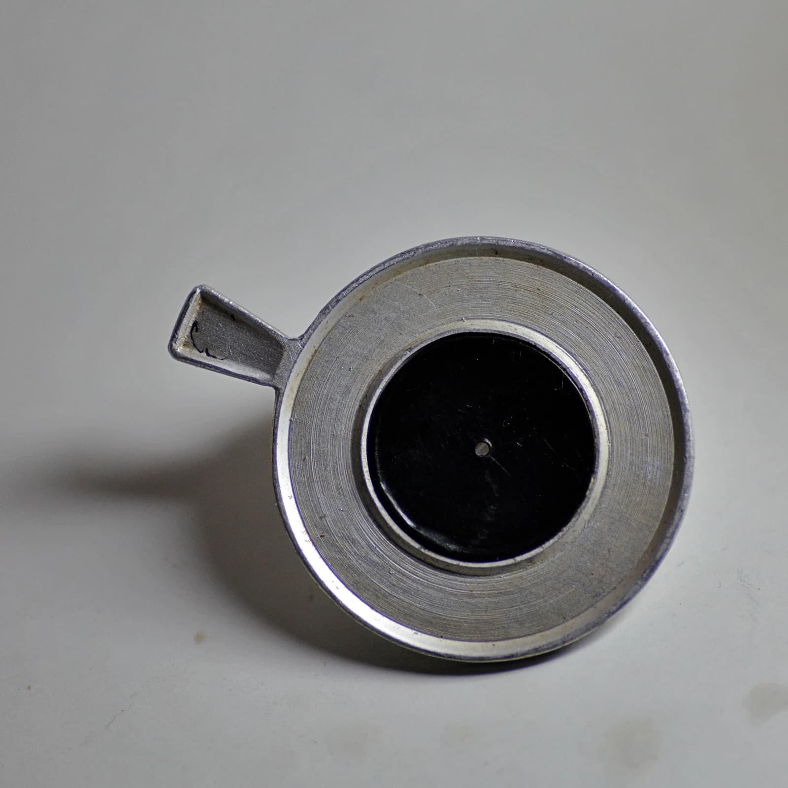
Pinhole occluder
Helps assess whether an optical refractive component may contribute to reduced visual acuity.
Identify · Locate · Practise 4 viva questions · Zoomable reference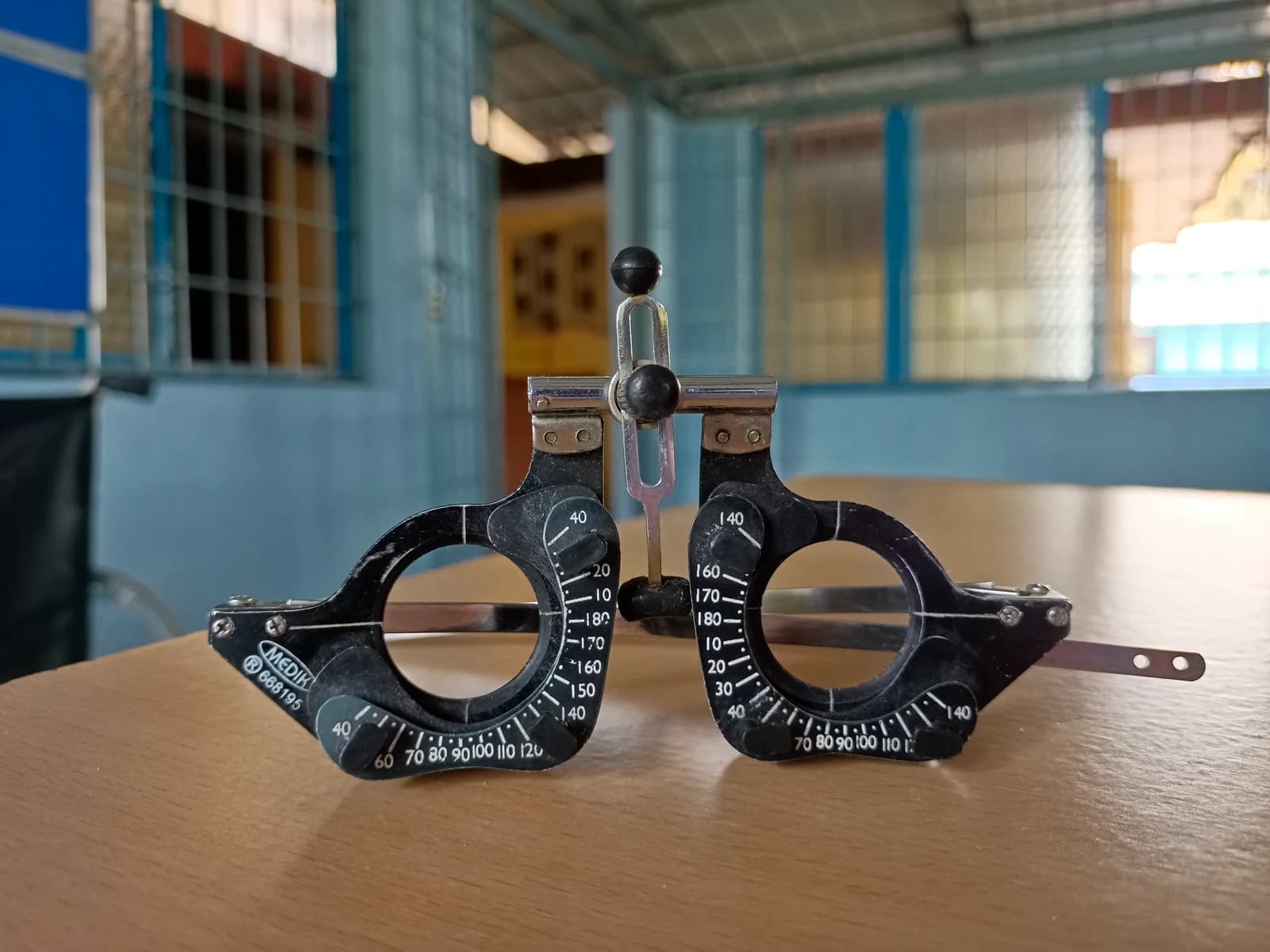
Ophthalmic trial frame
Holds trial lenses in front of the eyes during refraction and selected binocular-vision tests.
Identify · Locate · Practise 4 viva questions · Zoomable reference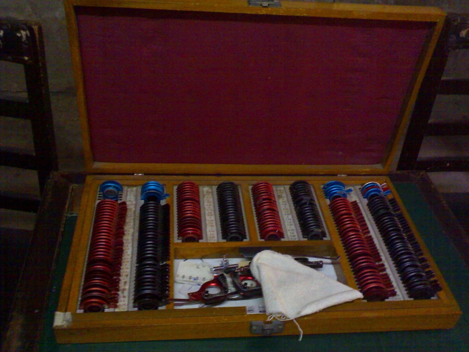
Trial lens set
Provides known optical elements for refraction and visual testing with a trial frame.
Identify · Locate · Practise 4 viva questions · Zoomable reference
Retinoscope
Provides objective assessment of refractive error by observation of the retinal reflex.
Identify · Locate · Practise 4 viva questions · Zoomable reference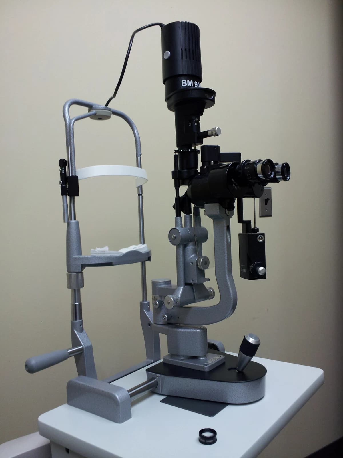
Slit-lamp biomicroscope
Provides magnified, illuminated examination of anterior eye structures and, with appropriate lenses, the posterior segment.
Identify · Locate · Practise 4 viva questions · Zoomable reference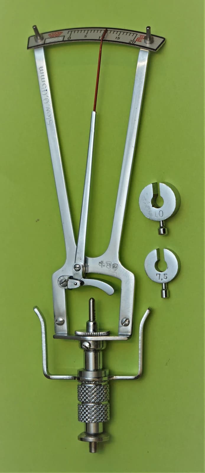
Schiøtz indentation tonometer
Estimates intraocular pressure by indentation in a supervised examination.
Identify · Locate · Practise 4 viva questions · Zoomable reference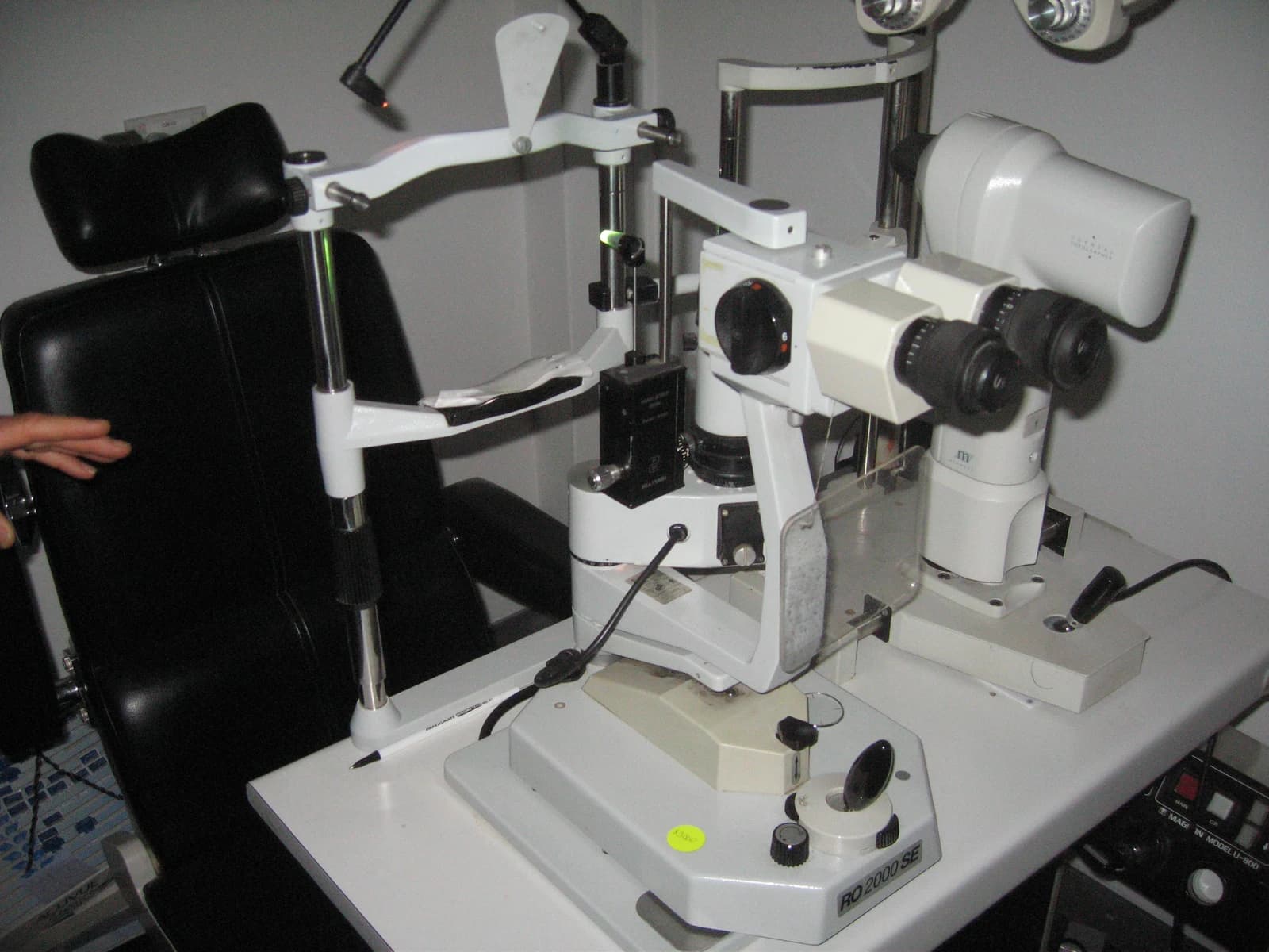
Applanation tonometer
Estimates intraocular pressure by flattening a defined corneal area.
Identify · Locate · Practise 4 viva questions · Zoomable reference
Lensmeter
Measures spectacle-lens power, cylinder axis and related optical properties.
Identify · Locate · Practise 4 viva questions · Zoomable reference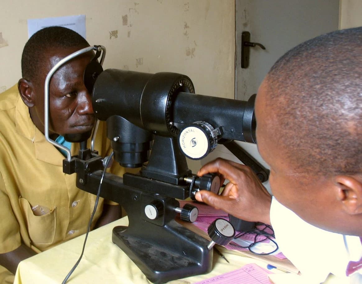
Keratometer
Estimates anterior corneal curvature and corneal astigmatism.
Identify · Locate · Practise 4 viva questions · Zoomable reference
Wire lid speculum
Keeps eyelids apart to provide exposure during an ophthalmic procedure.
Identify · Locate · Practise 4 viva questions · Zoomable reference
Universal eye speculum
Maintains lid separation during ophthalmic surgery.
Identify · Locate · Practise 4 viva questions · Zoomable reference
Barraquer needle holder
Holds an appropriate microsurgical needle during ophthalmic suturing.
Identify · Locate · Practise 4 viva questions · Zoomable reference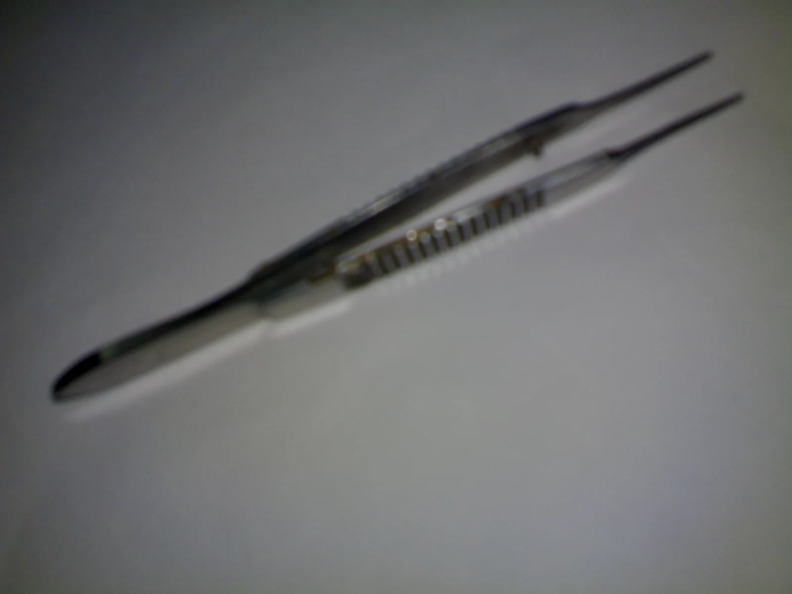
Suture-tying forceps
Grasps and manipulates fine suture material during microsurgical knot tying.
Identify · Locate · Practise 4 viva questions · Zoomable reference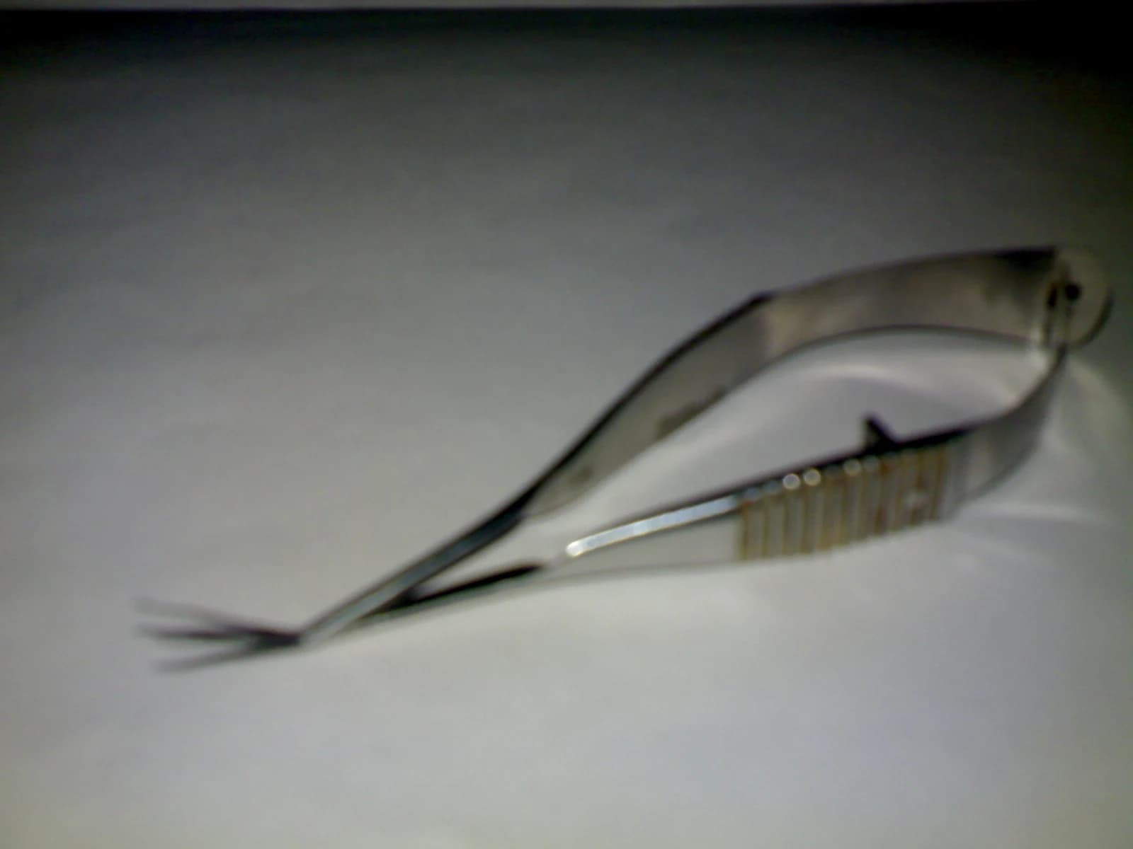
Vannas scissors
Makes delicate cuts in selected ophthalmic tissues under magnification.
Identify · Locate · Practise 4 viva questions · Zoomable reference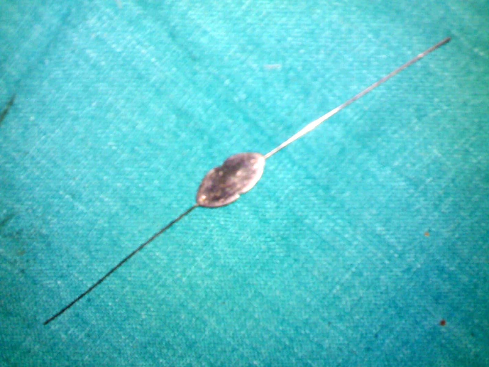
Bowman lacrimal probe
Assesses or traverses parts of the lacrimal drainage pathway in appropriately selected, supervised care.
Identify · Locate · Practise 4 viva questions · Zoomable reference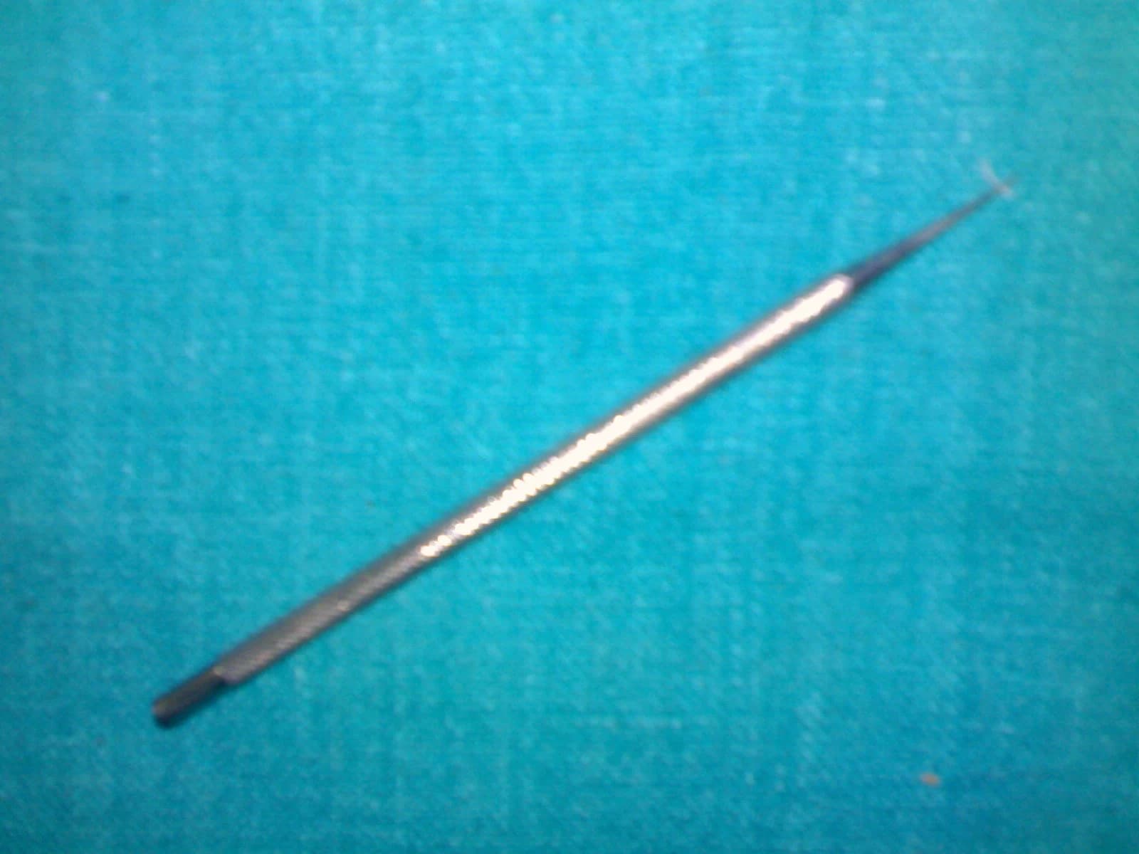
Punctum dilator
Enlarges the lacrimal punctal opening when clinically indicated before selected lacrimal procedures.
Identify · Locate · Practise 4 viva questions · Zoomable reference
Lacrimal irrigation cannula
Introduces fluid for lacrimal irrigation in an appropriate clinical assessment.
Identify · Locate · Practise 4 viva questions · Zoomable reference
Angular keratotome
Creates a controlled corneal or corneoscleral entry incision in selected ophthalmic surgery.
Identify · Locate · Practise 4 viva questions · Zoomable reference
Irrigation–aspiration cannula
Provides controlled fluid exchange and removal of selected material during ophthalmic surgery.
Identify · Locate · Practise 4 viva questions · Zoomable reference
Sinskey IOL hook
Manipulates an intraocular lens or its haptics during selected cataract procedures.
Identify · Locate · Practise 4 viva questions · Zoomable referenceA core MBBS teaching collection, grouped under third year for navigation. Exam timing varies by batch and university. Instrument models and practical lists vary by college. Photographs illustrate specific models; follow your department’s supervised practical programme.