 MEDIVEERSETHE PRACTICAL HUB
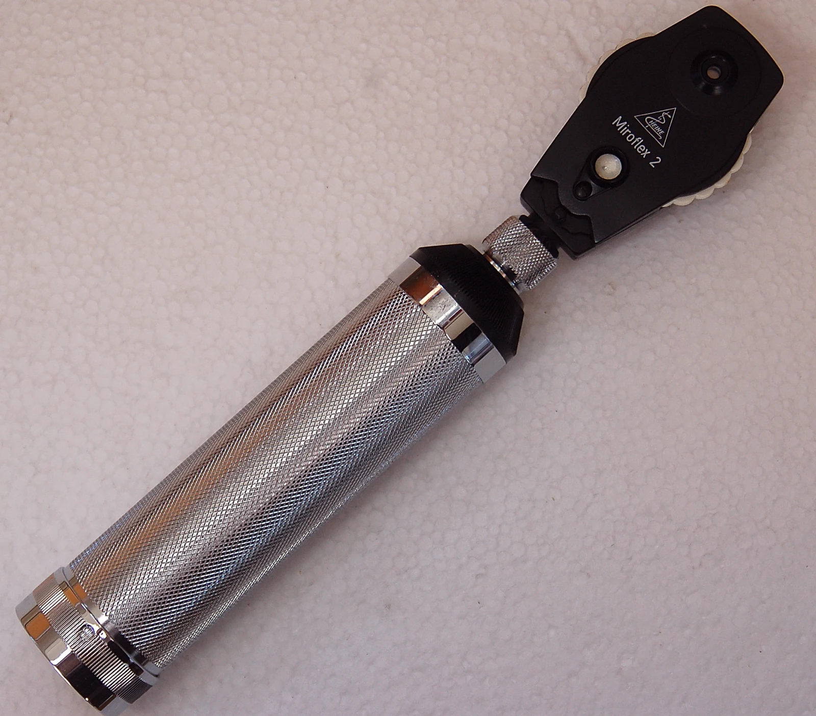
Viewing aperture: located on the reference instrument. Compare its shape and position with the apparatus used in your practical.
Zoom in, then drag to inspect. Keyboard: + / − to zoom, arrows to pan, 0 to reset. Tap the numbered markers to explore parts.
Direct ophthalmoscope — reference view
Image: Janee · Source / original · CC BY-SA 3.0Resized and converted to WebP; original colours retained.
Direct ophthalmoscope
Fundoscope · ophthalmoscope
What is its use?
Examines the ocular fundus, including the optic disc and retinal vessels.
Tap a number to locate its part. Zoom, drag or use full screen to inspect the reference. Models and graduations can differ; check the actual instrument used in your practical class or clinical posting.
How to recognise it.
- Illuminated head with a viewing aperture
- Lens-selection wheel
- Handle commonly contains the battery or power supply
An otoscope is designed for the ear canal and tympanic membrane, not the retinal fundus.
Point them out.
Choose a part to bring its numbered marker into view.
How it works.
Coaxial illumination and viewing optics allow light reflected from the fundus to reach the examiner; selectable lenses compensate for refractive differences.
Think first. Then reveal.
Explain your answer aloud before opening it.
01What structures are examined?+
The optic disc, retinal vessels and visible retina, with the field depending on the device and conditions.
02What does the lens wheel do?+
It changes lens power to assist focusing.
03What is the red reflex?+
The reddish reflection of light returning from within the eye.
04Does direct ophthalmoscopy provide a wide stereoscopic view?+
No. Conventional direct ophthalmoscopy gives a relatively narrow, magnified, non-stereoscopic view.
Read the references.
Teaching and clinical references are listed here. Image attribution and licence appear below the viewer.