 MEDIVEERSETHE PRACTICAL HUB
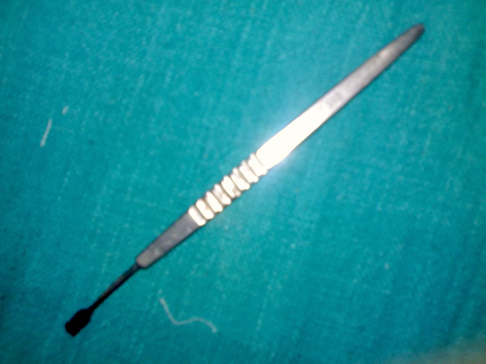
The small shaped cutting end creates the intended surgical entry; dimensions depend on the specific blade.
Zoom in, then drag to inspect. Keyboard: + / − to zoom, arrows to pan, 0 to reset. Tap the numbered markers to explore parts.
Angular keratotome — reference view
Image: Sarindam7 · Source / original · CC BY-SA 4.0Resized and converted to WebP; original colours retained.
Angular keratotome
Keratome · ophthalmic entry blade
What is its use?
Creates a controlled corneal or corneoscleral entry incision in selected ophthalmic surgery.
Tap a number to locate its part. Zoom, drag or use full screen to inspect the reference. Models and graduations can differ; check the actual instrument used in your practical class or clinical posting.
How to recognise it.
- Pointed angular blade
- Narrow shank
- Handle or hub depending on design
A keratome cuts an entry wound; a spatula or repositor mainly manipulates tissue without that cutting role.
Point them out.
Choose a part to bring its numbered marker into view.
How it works.
A shaped sharp blade cuts an entry of defined geometry; dimensions and bevel vary by model.
Think first. Then reveal.
Explain your answer aloud before opening it.
01What is its working part?+
The sharp shaped blade.
02Why does blade width matter?+
It influences incision dimensions and compatibility with the planned surgical system.
03Is every keratome identical?+
No. Width, angle, bevel and intended incision design vary.
04Is it a routine bedside examination instrument?+
No. It is a surgical cutting instrument requiring an appropriate operative setting.
Read the references.
Teaching and clinical references are listed here. Image attribution and licence appear below the viewer.