 MEDIVEERSETHE PRACTICAL HUB
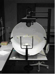
Test bowl: located on the reference instrument. Compare its shape and position with the apparatus used in your practical.
Zoom in, then drag to inspect. Keyboard: + / − to zoom, arrows to pan, 0 to reset. Tap the numbered markers to explore parts.
Visual-field perimeter — reference view
Image: Douwebergsma at Dutch Wikipedia · Source / original · CC BY-SA 3.0Resized and converted to WebP; original colours retained.
Visual-field perimeter
Perimetry apparatus · visual field tester
What is its use?
Maps the extent and sensitivity of the visual field.
Tap a number to locate its part. Zoom, drag or use full screen to inspect the reference. Models and graduations can differ; check the actual instrument used in your practical class or clinical posting.
How to recognise it.
- A fixation target and test field
- A support positions the head or eye
- A moving target or controlled light stimuli are presented
A Snellen chart measures central visual acuity; perimetry tests the visual field.
Point them out.
Choose a part to bring its numbered marker into view.
How it works.
Targets are presented at known locations while fixation is maintained. Kinetic and static perimetry use different presentation strategies.
Think first. Then reveal.
Explain your answer aloud before opening it.
01What is an isopter?+
A contour joining locations with the same visual sensitivity for a defined target.
02What is the physiological blind spot?+
The visual-field location corresponding to the optic disc, where photoreceptors are absent.
03Why maintain fixation?+
Eye movement changes the retinal location tested and makes the field map unreliable.
04How do static and kinetic perimetry differ?+
Static perimetry changes stimulus intensity at fixed locations; kinetic perimetry uses moving stimuli of defined characteristics.
Read the references.
Teaching and clinical references are listed here. Image attribution and licence appear below the viewer.