 MEDIVEERSETHE PRACTICAL HUB
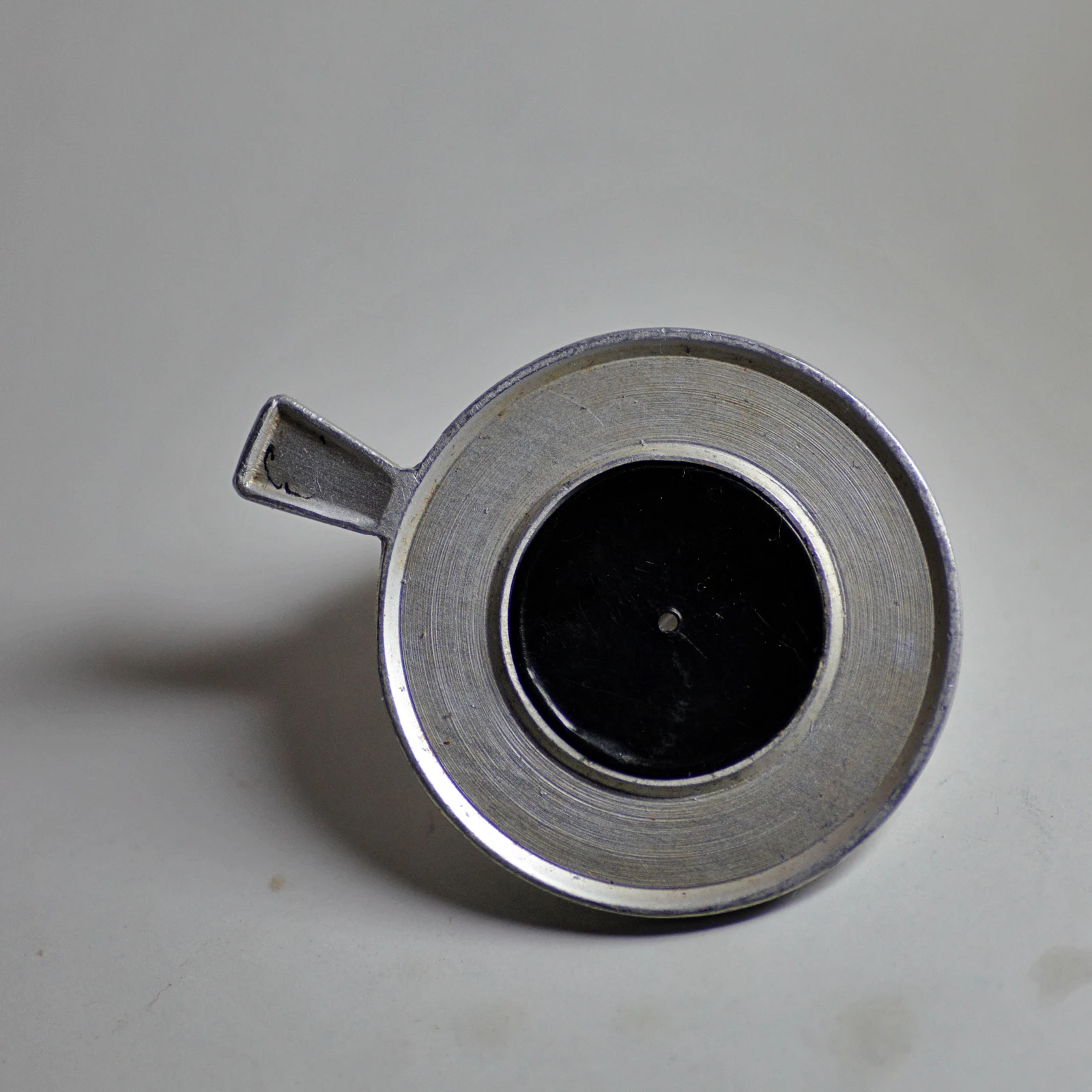
The small opening limits peripheral rays and can reduce defocus blur.
Zoom in, then drag to inspect. Keyboard: + / − to zoom, arrows to pan, 0 to reset. Tap the numbered markers to explore parts.
Pinhole occluder — reference view
Image: Ajeeshkumar4u · Source / original · CC BY-SA 4.0Resized and converted to WebP; original colours retained.
Pinhole occluder
Pinhole disc · acuity screening
What is its use?
Helps assess whether an optical refractive component may contribute to reduced visual acuity.
Tap a number to locate its part. Zoom, drag or use full screen to inspect the reference. Models and graduations can differ; check the actual instrument used in your practical class or clinical posting.
How to recognise it.
- Opaque trial-frame accessory
- Small central aperture
- Metal rim with a mounting tab
Improvement suggests an optical component; it does not by itself exclude disease or give a spectacle prescription.
Point them out.
Choose a part to bring its numbered marker into view.
How it works.
Restricting peripheral rays reduces the retinal blur circle and increases depth of focus, within the aperture’s optical limits.
Think first. Then reveal.
Explain your answer aloud before opening it.
01Why can acuity improve through a pinhole?+
A smaller bundle of rays reduces the size of defocus blur on the retina.
02Does no improvement prove retinal disease?+
No. Several ocular and testing factors can limit improvement; interpret with the rest of the examination.
03Can the aperture be arbitrarily small?+
No. Very small apertures reduce light and introduce diffraction.
04Does this measure refractive power?+
No. Objective and subjective refraction are needed to establish a suitable correction.
Read the references.
Teaching and clinical references are listed here. Image attribution and licence appear below the viewer.