 MEDIVEERSETHE PRACTICAL HUB
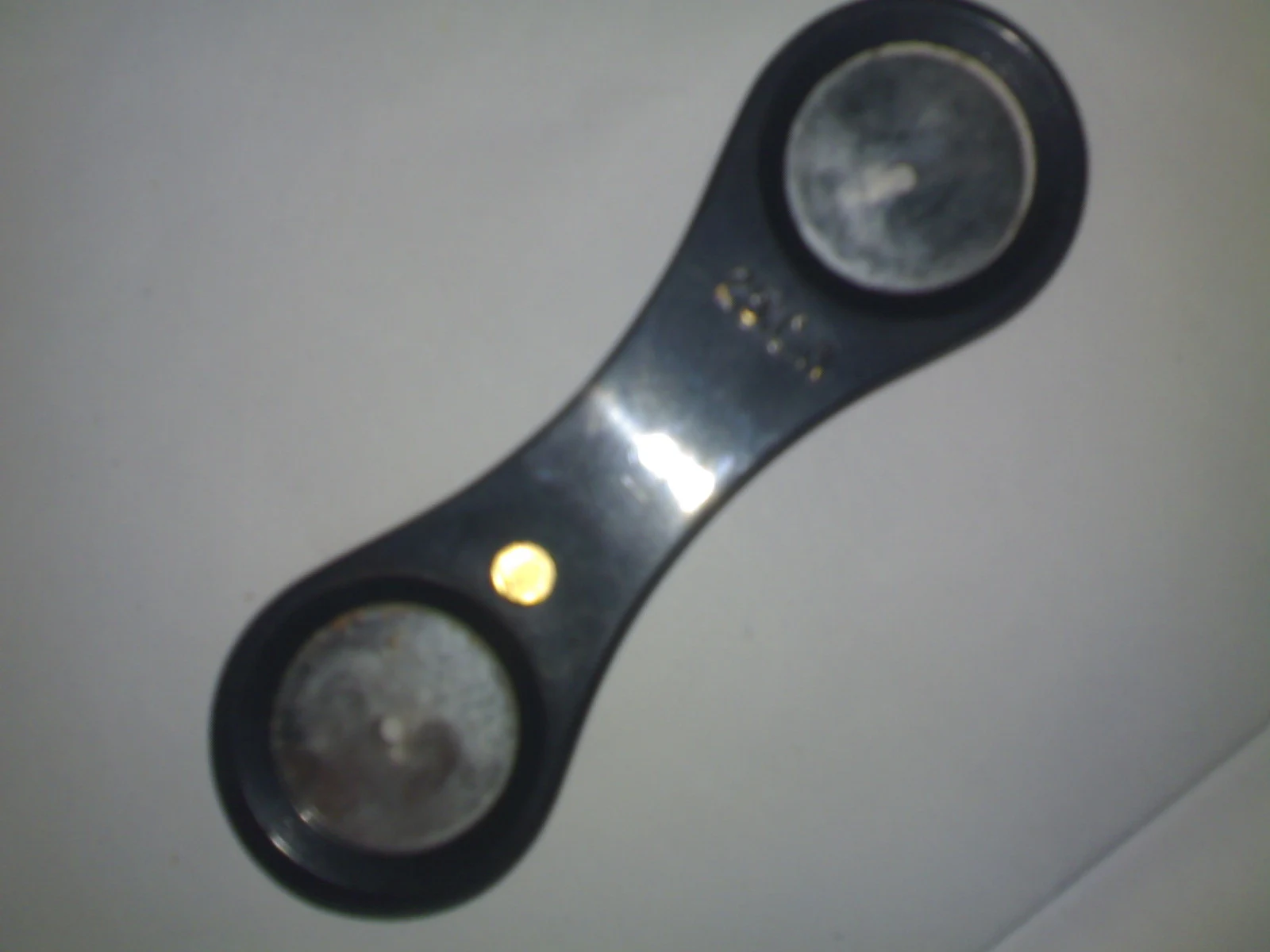
A mirror face on this historical mirror retinoscope. Modern streak retinoscopes use an illuminated head instead.
Zoom in, then drag to inspect. Keyboard: + / − to zoom, arrows to pan, 0 to reset. Tap the numbered markers to explore parts.
Historical mirror retinoscope — reference view
Image: Sarindam7 · Source / original · CC BY-SA 3.0Resized and converted to WebP; original colours retained.
Retinoscope
Retinoscopy mirror · streak retinoscope
What is its use?
Provides objective assessment of refractive error by observation of the retinal reflex.
Tap a number to locate its part. Zoom, drag or use full screen to inspect the reference. Models and graduations can differ; check the actual instrument used in your practical class or clinical posting.
How to recognise it.
- Two mirror faces joined in a handheld body on this historical reference
- Mirror-based observation rather than a modern electric streak head
- Inspect mirror curvature and viewing aperture on the actual instrument
A direct ophthalmoscope is primarily used to inspect fundus detail; a retinoscope is primarily used for objective refraction. The reference photograph shows a historical mirror retinoscope, not a modern battery-powered streak model.
Point them out.
Choose a part to bring its numbered marker into view.
How it works.
The examiner interprets reflex movement and neutralisation using known lenses, accounting for working distance.
Think first. Then reveal.
Explain your answer aloud before opening it.
01What is neutralisation?+
The reflex fills the pupil without an apparent directional movement in the testing configuration.
02Why account for working distance?+
The examination is performed at a finite distance; its vergence contribution must be removed from the gross finding.
03Does retinoscopy require the patient to compare lenses verbally?+
Not for the objective observation, although fixation, accommodation and cooperation still matter.
04Why does a streak retinoscope help with astigmatism?+
Its oriented beam helps assess the principal meridians and the cylinder axis.
Read the references.
Teaching and clinical references are listed here. Image attribution and licence appear below the viewer.