 MEDIVEERSETHE PRACTICAL HUB
Provide binocular viewing through the microscope.
Zoom in, then drag to inspect. Keyboard: + / − to zoom, arrows to pan, 0 to reset. Tap the numbered markers to explore parts.
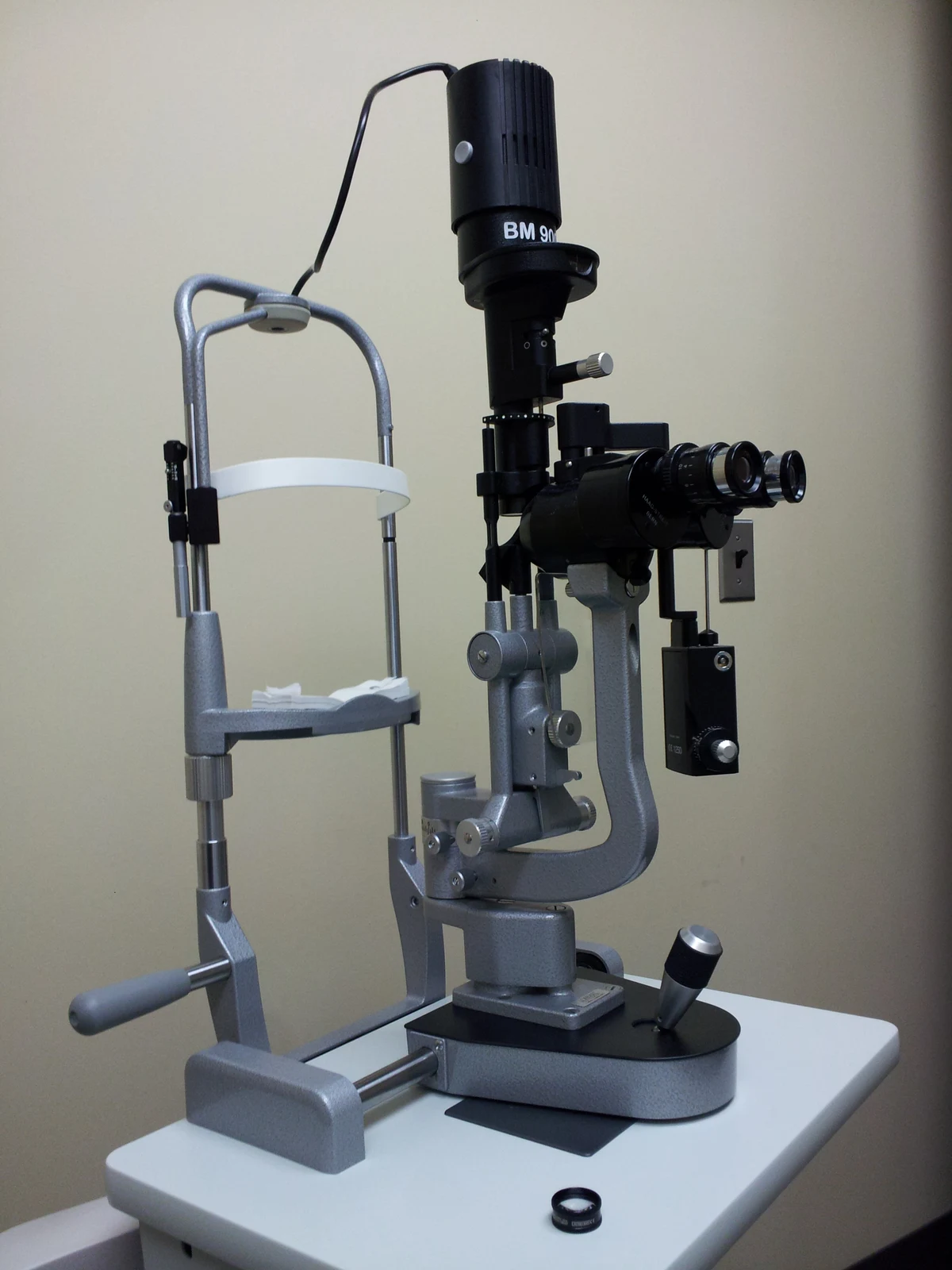
Slit-lamp biomicroscope — reference view
Image: Ben Schumin · Source / original · CC BY-SA 3.0Resized and converted to WebP; original colours retained.
Slit-lamp biomicroscope
Slit lamp · biomicroscope
What is its use?
Provides magnified, illuminated examination of anterior eye structures and, with appropriate lenses, the posterior segment.
Tap a number to locate its part. Zoom, drag or use full screen to inspect the reference. Models and graduations can differ; check the actual instrument used in your practical class or clinical posting.
How to recognise it.
- Binocular observation microscope
- Adjustable illumination arm
- Chin and forehead supports
A slit lamp reveals ocular structures; tonometry is a separate pressure-measurement function, sometimes mounted on it.
Point them out.
Choose a part to bring its numbered marker into view.
How it works.
A narrow adjustable beam creates an optical section while the microscope provides stereoscopic magnification.
Think first. Then reveal.
Explain your answer aloud before opening it.
01Why use a slit beam?+
It provides optical sectioning and helps judge tissue depth and location.
02Why stabilise the head?+
Steady alignment improves focus and control during examination.
03Can the posterior segment be seen?+
Yes, with suitable examination lenses and clinical technique; the basic instrument alone is not a direct ophthalmoscope.
04Is it only for the cornea?+
No. The lids, conjunctiva, cornea, anterior chamber, iris and lens are also assessed, with additional techniques extending the view.
Read the references.
Teaching and clinical references are listed here. Image attribution and licence appear below the viewer.