 MEDIVEERSETHE PRACTICAL HUB
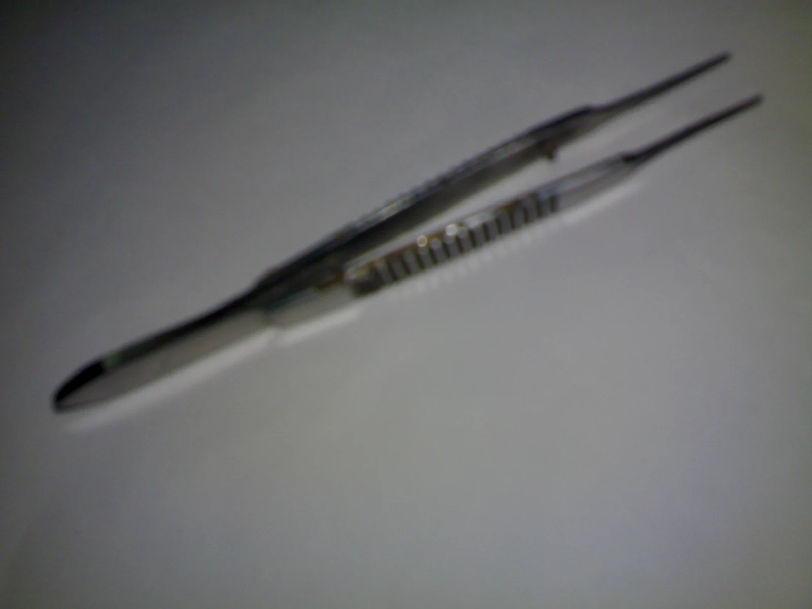
Fine opposed ends manipulate suture; the microscopic tying surface is not resolved in this wide reference.
Zoom in, then drag to inspect. Keyboard: + / − to zoom, arrows to pan, 0 to reset. Tap the numbered markers to explore parts.
Suture-tying forceps — reference view
Image: Sarindam7 · Source / original · CC BY-SA 3.0Resized and converted to WebP; original colours retained.
Suture-tying forceps
Ophthalmic tying forceps
What is its use?
Grasps and manipulates fine suture material during microsurgical knot tying.
Tap a number to locate its part. Zoom, drag or use full screen to inspect the reference. Models and graduations can differ; check the actual instrument used in your practical class or clinical posting.
How to recognise it.
- Fine opposed tips
- Spring handle
- Smooth tying platform on suitable models
Toothed fixation forceps grip tissue; tying forceps are intended for handling fine suture.
Point them out.
Choose a part to bring its numbered marker into view.
How it works.
Fine controlled apposition of the tips holds suture without the broad crush of a heavier clamp.
Think first. Then reveal.
Explain your answer aloud before opening it.
01Why avoid crushing suture?+
Damage can weaken the material and compromise the knot or wound closure.
02What is the tying platform?+
A small smooth working surface near the tips used to hold suture.
03Are all eye forceps interchangeable?+
No. Tip design determines whether the instrument is suited to tissue, suture or another task.
04Why use magnification for fine work?+
It improves visual control of small structures and instrument tips.
Read the references.
Teaching and clinical references are listed here. Image attribution and licence appear below the viewer.