 MEDIVEERSETHE PRACTICAL HUB
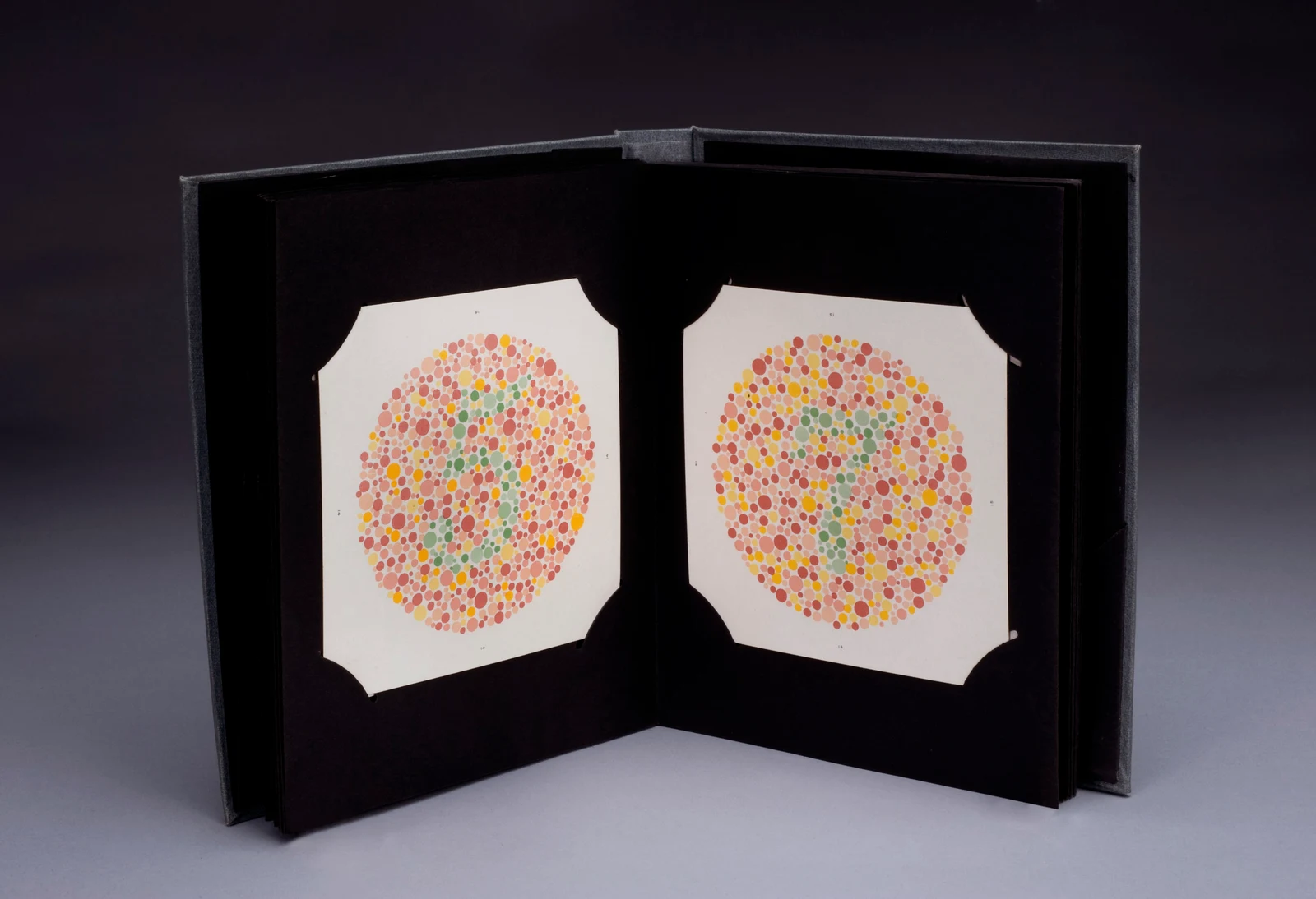
Coloured-dot plate: located on the reference instrument. Compare its shape and position with the apparatus used in your practical.
Zoom in, then drag to inspect. Keyboard: + / − to zoom, arrows to pan, 0 to reset. Tap the numbered markers to explore parts.
Ishihara colour-vision plates — reference view
Image: https://wellcomeimages.org/indexplus/obf_images/2b/03/98b13461c74c9d886b8367aeb439.jpg Gallery: https://wellcomeimages.org/indexplus/image/L0058975.html Wellcome Collection gallery (2018-03-30): https://wellcomecollection.org/works/bycpde6e CC-BY-4.0 · Source / original · CC BY 4.0Resized and converted to WebP; original colours retained.
Ishihara colour-vision plates
Pseudoisochromatic plates · colour vision test
What is its use?
Screens principally for red–green colour-vision deficiency.
Tap a number to locate its part. Zoom, drag or use full screen to inspect the reference. Models and graduations can differ; check the actual apparatus used in your laboratory.
How to recognise it.
- Patterns made from many coloured dots
- A numeral or path is embedded in the pattern
- Plates are presented in a standard sequence under suitable illumination
Ishihara plates do not comprehensively classify every acquired or blue–yellow colour-vision problem. A screen image is not a calibrated clinical test.
Point them out.
Choose a part to bring its numbered marker into view.
The science behind the reading.
The embedded figure and surrounding dots differ in ways that depend on colour discrimination rather than just reading ability.
Think first. Then reveal.
Explain your answer aloud before opening it.
01What deficiency is commonly screened?+
Red–green colour-vision deficiency.
02Why are pseudoisochromatic dots used?+
They form an embedded figure that may be difficult to distinguish when the relevant colour signals are confused.
03Why does illumination matter?+
The appearance of the colours changes with lighting and reproduction.
04Can a website image diagnose colour deficiency?+
No. Displays and reproduction alter colour; formal testing requires suitable validated materials and conditions.
Read the references.
Teaching and clinical references are listed here. Image attribution and licence appear below the viewer.