 MEDIVEERSETHE PRACTICAL HUB
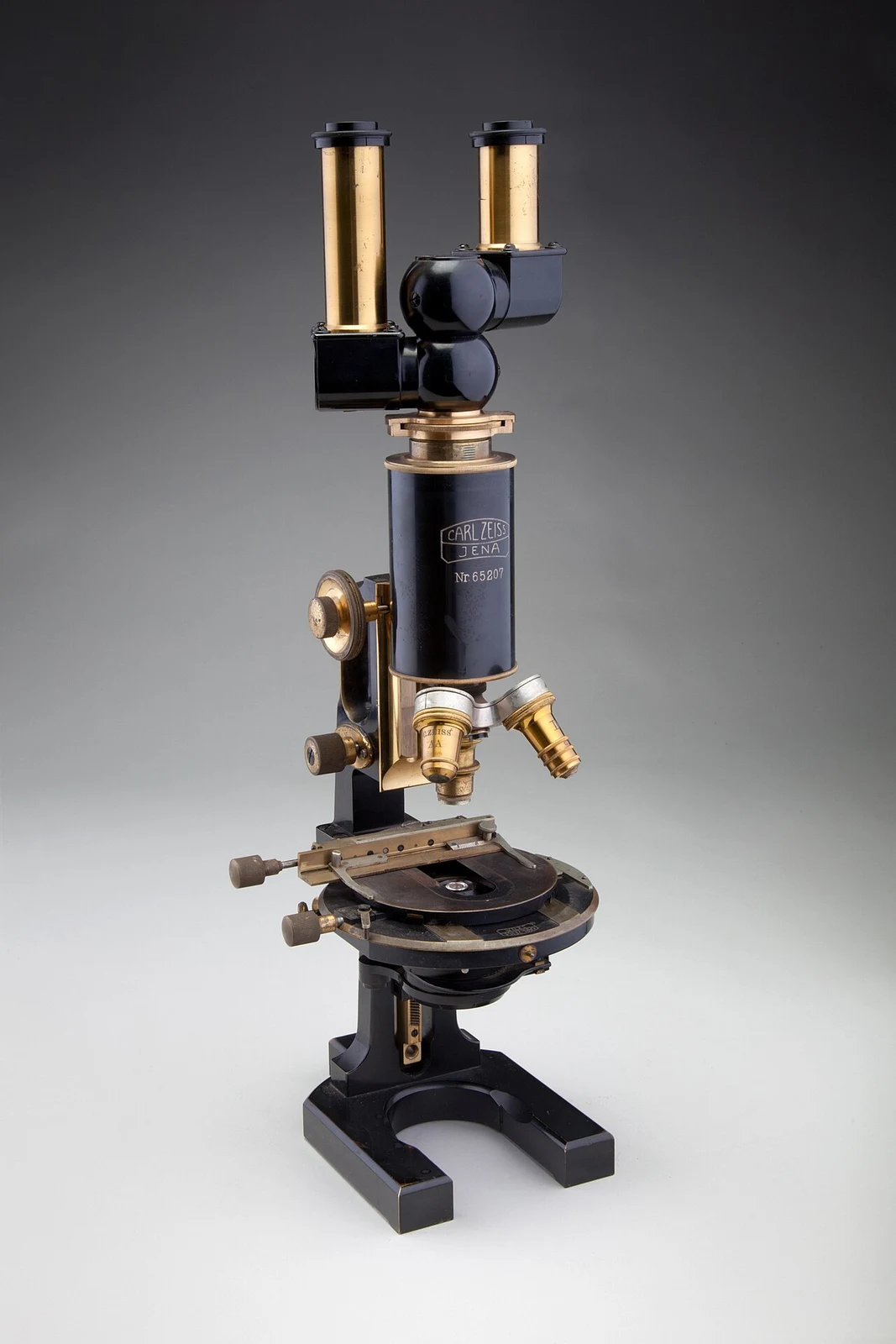
Eyepiece: located on the reference instrument. Compare its shape and position with the apparatus used in your practical.
Zoom in, then drag to inspect. Keyboard: + / − to zoom, arrows to pan, 0 to reset. Tap the numbered markers to explore parts.
Compound microscope — reference view
Image: Chad Anderson, staff photographer for SFO Museum · Source / original · CC BY-SA 2.0Resized and converted to WebP; original colours retained.
Compound microscope
Light microscope · optical microscope
What is its use?
Magnifies cells and tissues for practical identification and manual counting.
Tap a number to locate its part. Zoom, drag or use full screen to inspect the reference. Models and graduations can differ; check the actual apparatus used in your laboratory.
How to recognise it.
- Eyepiece above a revolving objective turret
- Mechanical stage holds the slide
- Coarse and fine focus controls move the focusing mechanism
A stereomicroscope provides a lower-magnification three-dimensional view; a compound microscope is used for transmitted-light examination of thin specimens.
Point them out.
Choose a part to bring its numbered marker into view.
The science behind the reading.
The objective forms a magnified intermediate image; the eyepiece magnifies it further. Total magnification is objective × eyepiece magnification.
Think first. Then reveal.
Explain your answer aloud before opening it.
01How is total magnification calculated?+
Multiply objective magnification by eyepiece magnification; 40× and 10× give 400×.
02Why use fine rather than coarse focus at high power?+
Fine focus gives controlled small adjustments and reduces the chance of driving an objective into the slide.
03What is resolving power?+
The ability to distinguish two closely spaced points as separate; magnification alone does not improve resolution.
04Why use immersion oil with a designated oil objective?+
It reduces refractive mismatch between glass and the objective and permits a higher numerical aperture. Use only objectives intended for oil.
Read the references.
Teaching and clinical references are listed here. Image attribution and licence appear below the viewer.