 MEDIVEERSETHE PRACTICAL HUB
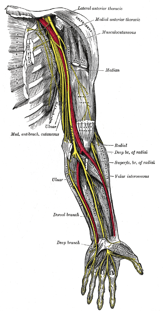
Ulnar nerve leaves the anterior arm neurovascular axis toward the medial elbow.
Zoom in, then drag to inspect. Keyboard: + / − to zoom, arrows to pan, 0 to reset. Tap the numbered markers to explore parts.
upper nerves: original labelled anatomical illustration.
Image: Henry Vandyke Carter / Gray’s Anatomy (1918) · Source / original · Public domainNumbered markers and focused views are Mediveerse learning overlays; the original reference is linked above.
Ulnar nerve
Understand what you see.
Supplies most intrinsic hand muscles and sensation over the medial hand.
Zoom into the original labels. Practise pointing out each structure, then explain it aloud. Diagrams are teaching references; specimen appearance and slide staining vary.
How to recognise it.
- Medial cord terminal branch
- Passes behind medial epicondyle
- Enters hand superficial to flexor retinaculum through Guyon canal
Point them out.
Tap a number to open its reference, zoom in and locate the structure. Numbers stay the same across views. This key follows landmarks visible in the reference images.
Think first. Then reveal.
Say your answer aloud before opening the suggested response.
01How do you identify ulnar nerve?+
Medial cord terminal branch. Passes behind medial epicondyle. Enters hand superficial to flexor retinaculum through Guyon canal.
02Which lumbricals does it supply?+
Usually the third and fourth through its deep branch.
03Is it inside the carpal tunnel?+
No. It passes in Guyon canal, superficial to flexor retinaculum.
04What is its main functional or developmental significance?+
Supplies most intrinsic hand muscles and sensation over the medial hand. Key structures to recognise: Flexor carpi ulnaris supply; Deep hand branch; Superficial sensory branch.
Read the references.
Credited anatomical plates, microscopy and university teaching references. Individual image credits appear below the viewer. Historical plates can use older terminology; use the lesson text for current names.