Identify the pattern
Compare deeper cells with flattened superficial cells in this coloured teaching schematic.
 MEDIVEERSETHE PRACTICAL HUB
OBSERVE / REASON / EXPLAIN
Recognise the stratified squamous pattern and relate epithelial variation to oral mucosal sites.
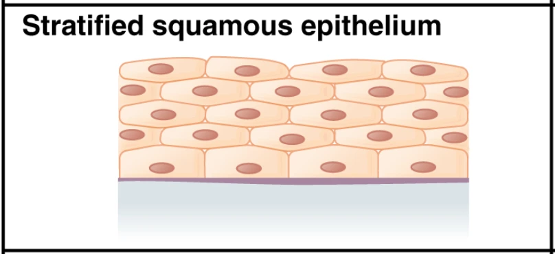
Flattened cells at the free epithelial surface in this schematic.
Zoom in, then drag to inspect. Keyboard: + / − to zoom, arrows to pan, 0 to reset. Tap the numbered markers to explore parts.
Stratified squamous epithelium — coloured teaching schematic
Image: OpenStax / University of Hawaiʻi adapted teaching figures; original contributors credited at source · Source / original · CC BY 4.0 — figure credits at sourceResized and converted to WebP; original colours retained.
Stratified squamous epithelium has multiple cell layers with flattened surface cells. Oral sites vary in keratinisation and underlying tissue arrangement; this reference is a schematic, not a patient micrograph.
Compare deeper cells with flattened superficial cells in this coloured teaching schematic.
Discuss lining, masticatory and specialised oral mucosa; do not infer every site-specific feature from this generic diagram.
Use tissue orientation, surface keratinisation and the underlying connective tissue in an authorised histology specimen.
Try answering aloud, then open the suggested answer. Examiner questions vary.
By its multiple layers and the flattened shape of the superficial cells.
No. Keratinisation varies by anatomical site and mucosal type.
No. It illustrates a general epithelial pattern and is not a diagnostic micrograph.
It helps orient the tissue and identify its structural relationship with the underlying connective tissue.