Observe
Identify the visible mineralised developing crowns. This is a radiograph, not a histological section.
 MEDIVEERSETHE PRACTICAL HUB
OBSERVE / REASON / EXPLAIN
Recognise developing molar structures on a teaching radiograph and distinguish formation from eruption.
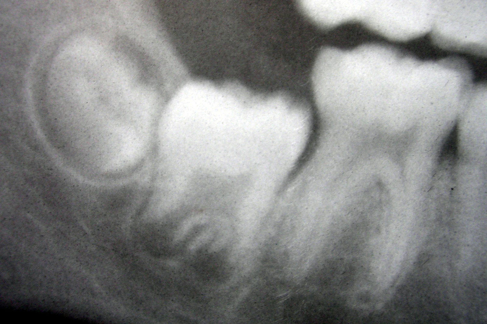
A mineralised developing crown in the teaching radiograph.
Zoom in, then drag to inspect. Keyboard: + / − to zoom, arrows to pan, 0 to reset. Tap the numbered markers to explore parts.
Developing molars — radiographic reference
Image: No machine-readable author provided. Dozenist assumed (based on copyright claims). · Source / original · CC BY-SA 3.0Resized and converted to WebP; original colours retained.
Tooth development involves coordinated epithelial and ectomesenchymal interactions followed by hard-tissue formation.
Identify the visible mineralised developing crowns. This is a radiograph, not a histological section.
Distinguish developmental stages from eruption chronology.
Relate enamel and dentine formation to their responsible cells.
Try answering aloud, then open the suggested answer. Examiner questions vary.
Ameloblasts.
Odontoblasts.
No. Development and movement into the oral cavity are related but distinct processes.
No. Histological staging requires suitable sections and context.