Orient
Distinguish crown, neck and root before naming tissues.
 MEDIVEERSETHE PRACTICAL HUB
OBSERVE / REASON / EXPLAIN
Identify the principal tissues of a tooth on a coloured anatomical reference.
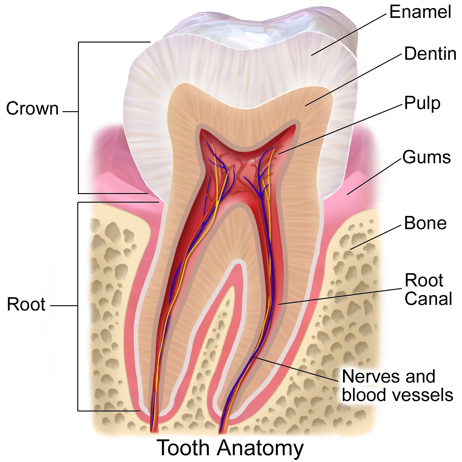
The outer mineralised layer covering the anatomical crown.
Zoom in, then drag to inspect. Keyboard: + / − to zoom, arrows to pan, 0 to reset. Tap the numbered markers to explore parts.
Tooth anatomy — coloured teaching schematic
Image: BruceBlaus. When using this image in external sources it can be cited as: Blausen.com staff (2014). "Medical gallery of Blausen Medical 2014". WikiJournal of Medicine 1 (2). DOI:10.15347/wjm/2014.010. ISSN 2002-4436. · Source / original · CC BY 3.0Resized and converted to WebP; original colours retained.
Enamel covers the crown, dentine forms most of the tooth, and pulp occupies the central cavity; cementum covers the root.
Distinguish crown, neck and root before naming tissues.
Compare enamel, dentine, pulp and surrounding supporting structures.
Explain tissue function without treating a schematic as a histological slide.
Try answering aloud, then open the suggested answer. Examiner questions vary.
Enamel.
No. It is vascular connective tissue containing nerves.
Cementum.
No. It simplifies anatomy for teaching.