 MEDIVEERSETHE PRACTICAL HUB
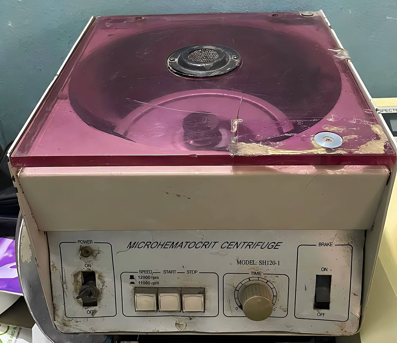
Lid: located on the reference instrument. Compare its shape and position with the apparatus used in your practical.
Zoom in, then drag to inspect. Keyboard: + / − to zoom, arrows to pan, 0 to reset. Tap the numbered markers to explore parts.
Microhaematocrit centrifuge — reference view
Image: Confidence24 · Source / original · CC BY-SA 4.0Resized and converted to WebP; original colours retained.
Microhaematocrit centrifuge
Capillary centrifuge · PCV centrifuge
What is its use?
Separates blood in capillary tubes for packed cell volume measurement.
Tap a number to locate its part. Zoom, drag or use full screen to inspect the reference. Models and graduations can differ; check the actual apparatus used in your laboratory.
How to recognise it.
- Rotor accommodates sealed capillary tubes
- Protective lid encloses the rotating assembly
- Controls specify operating conditions
A general-purpose centrifuge may use a different rotor and tubes. RPM alone does not specify the same force for different rotor radii.
Point them out.
Choose a part to bring its numbered marker into view.
The science behind the reading.
Centrifugal acceleration separates components according to their sedimentation behaviour. Packed red-cell length is compared with total sample length.
Think first. Then reveal.
Explain your answer aloud before opening it.
01How is PCV read?+
Packed red-cell column length divided by the total blood column length, excluding the buffy coat from the red-cell fraction.
02Why balance the rotor?+
Uneven loading causes vibration and can damage the instrument or tubes.
03What is the difference between RPM and RCF?+
RPM is rotational speed; RCF also depends on rotor radius and describes acceleration relative to gravity.
04Why seal a capillary end?+
The capillary must retain its sample during the validated centrifugation procedure.
Read the references.
Teaching and clinical references are listed here. Image attribution and licence appear below the viewer.