 MEDIVEERSETHE PRACTICAL HUB
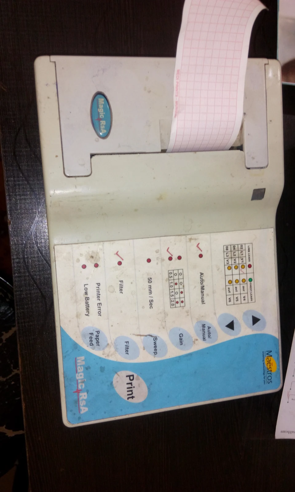
Paper trace: located on the reference instrument. Compare its shape and position with the apparatus used in your practical.
Zoom in, then drag to inspect. Keyboard: + / − to zoom, arrows to pan, 0 to reset. Tap the numbered markers to explore parts.
Electrocardiograph — reference view
Image: Zeeshan2907 · Source / original · CC BY-SA 4.0Resized and converted to WebP; original colours retained.
Electrocardiograph
ECG machine · EKG recorder
What is its use?
Records differences in electrical potential generated by cardiac activity at the body surface.
Tap a number to locate its part. Zoom, drag or use full screen to inspect the reference. Models and graduations can differ; check the actual apparatus used in your laboratory.
How to recognise it.
- Electronic acquisition unit with controls
- Display or paper output for waveforms
- Lead connections receive surface-electrode signals
An ECG records electrical activity, not contraction force or blood pressure. Ten electrodes provide the standard 12-lead ECG.
Point them out.
Choose a part to bring its numbered marker into view.
The science behind the reading.
Electrodes detect voltage differences; amplification and recording produce leads that view the cardiac electrical field from different directions.
Think first. Then reveal.
Explain your answer aloud before opening it.
01What does the P wave represent?+
Atrial depolarisation.
02What does the QRS complex represent?+
Ventricular depolarisation; atrial repolarisation is usually obscured within it.
03How many electrodes are used for a standard 12-lead ECG?+
Ten: four limb and six precordial electrodes. A lead is a view, not simply an electrode.
04What does the T wave represent?+
Ventricular repolarisation.
Read the references.
Teaching and clinical references are listed here. Image attribution and licence appear below the viewer.