 MEDIVEERSETHE PRACTICAL HUB
Head & Neck
Identify. Understand. Recall.
Skull, deep spaces, glands, pharynx, ear, nerves and vessels.
Open the full named checklistYour practical collection.
171 topics · Open a card to study its reference images and viva.
Every published topic, organised.
171 linked lessons. Open a group, choose a structure, then inspect its anatomical references and identification labels.
01Regional overviews5
Browse this group →02Skull bones12
Browse this group →03Sutures & surface landmarks8
Browse this group →04Foramina & canals15
Browse this group →05Nose, orbit & palate7
Browse this group →06Temporal bone landmarks5
Browse this group →07Mandible landmarks7
Browse this group →10Sphenoid landmarks4
Browse this group →11Ethmoid landmarks4
Browse this group →12Face & scalp muscles4
Browse this group →14Suprahyoid & infrahyoid muscles8
Browse this group →16Deep spaces10
Browse this group →17Salivary glands & ducts6
Browse this group →18Pharynx & palate8
Browse this group →19Larynx6
Browse this group →20Ear13
Browse this group →21Nerves & ganglia26
Browse this group →- Olfactory nerve (I)
- Optic nerve (II)
- Oculomotor nerve (III)
- Trochlear nerve (IV)
- Ophthalmic division (V1)
- Maxillary division (V2)
- Mandibular division (V3)
- Abducens nerve (VI)
- Facial nerve (VII)
- Vestibulocochlear nerve (VIII)
- Glossopharyngeal nerve (IX)
- Vagus nerve (X)
- Accessory nerve (XI)
- Hypoglossal nerve (XII)
- Lingual nerve
- Inferior alveolar nerve
- Auriculotemporal nerve
- Chorda tympani
- Superior laryngeal nerve
- Recurrent laryngeal nerve
- Ansa cervicalis
- Cervical plexus
- Cervical sympathetic trunk
- Otic ganglion
- Pterygopalatine ganglion
- Submandibular ganglion
22Vessels & lymphatics16
Browse this group →- Common carotid artery
- External carotid artery
- Internal carotid artery
- Carotid sinus and body
- Facial artery
- Lingual artery
- Maxillary artery
- Middle meningeal artery
- Superficial temporal artery
- Vertebral artery
- Internal jugular vein
- External jugular vein
- Retromandibular vein
- Pterygoid venous plexus
- Cavernous sinus
- Deep cervical lymph nodes
Includes deep spaces, salivary glands, pharynx and larynx, ear, nerves, vessels and lymphatics. This undergraduate revision collection supports your departmental syllabus; anatomical variants and specimen appearances differ. Galleries contain anatomical plates and illustrations with original labels and individual image credits.

Skull: views & sutures
Protects the brain and supports facial and sensory structures.
Identify · Structures · Viva Focused view + full reference · 6 viva prompts
Mandible
Supports mandibular teeth and moves during chewing and speech.
Identify · Structures · Viva 2 references · 5 viva prompts
Maxilla
Supports upper teeth and contributes to the orbit, nasal cavity and hard palate.
Identify · Structures · Viva 2 references · 5 viva prompts
Temporal bone
Houses ear structures and contributes to the temporomandibular joint.
Identify · Structures · Viva 2 references · 5 viva prompts
Sphenoid bone
Contributes to the cranial floor, orbit and passages for nerves and vessels.
Identify · Structures · Viva 2 references · 5 viva prompts
Cranial fossae & skull base
Supports brain regions and transmits cranial nerves and vessels.
Identify · Structures · Viva 2 references · 5 viva prompts
Hyoid bone
Anchors muscles involved in tongue movement and swallowing.
Identify · Structures · Viva 1 reference · 5 viva prompts
Muscles of mastication
Move the mandible during mastication.
Identify · Structures · Viva 1 reference · 5 viva prompts
Tongue: extrinsic muscles
Extrinsic muscles position the tongue during speech and swallowing.
Identify · Structures · Viva 1 reference · 5 viva promptsFrontal bone
Forms the forehead and roof of the orbit.
Identify · Structures · Viva Focused view + full reference · 3 viva promptsCoronal suture
A serrated junction between frontal and parietal bones.
Identify · Structures · Viva Focused view + full reference · 3 viva promptsGlabella
Smooth midline area of frontal bone between the superciliary arches.
Identify · Structures · Viva Focused view + full reference · 3 viva promptsSupraorbital foramen / notch
An opening or notch on the superior orbital rim.
Identify · Structures · Viva Focused view + full reference · 3 viva promptsSupraorbital margin
The superior rim of the bony orbit.
Identify · Structures · Viva Focused view + full reference · 3 viva prompts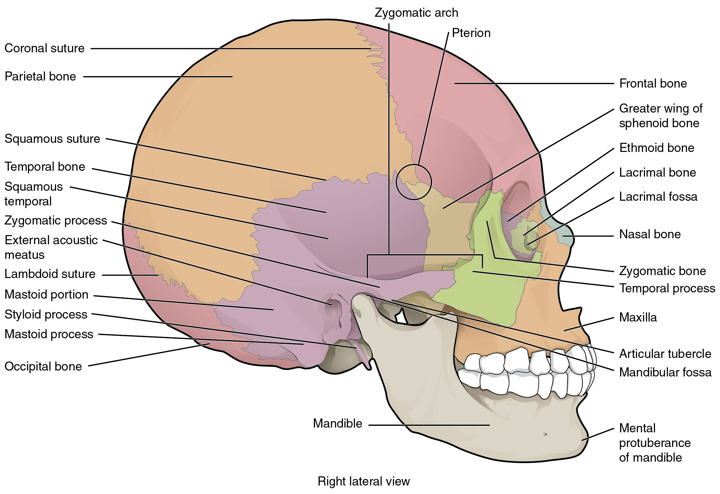
Parietal bone
Paired vault bones superior to the squamous temporal bone.
Identify · Structures · Viva Focused view + full reference · 3 viva promptsOccipital bone
Forms the posterior skull and much of the posterior cranial floor.
Identify · Structures · Viva Focused view + full reference · 3 viva promptsNasal bone
A paired small bone at the nasal bridge.
Identify · Structures · Viva Focused view + full reference · 3 viva promptsZygomatic bone
Forms the cheek prominence.
Identify · Structures · Viva Focused view + full reference · 3 viva promptsLacrimal bone
A small thin bone in the medial orbital wall.
Identify · Structures · Viva Focused view + full reference · 3 viva promptsVomer
Thin midline bone forming the posteroinferior bony nasal septum.
Identify · Structures · Viva Focused view + full reference · 3 viva promptsInferior nasal concha
A separate curved bone on the lateral nasal wall.
Identify · Structures · Viva Focused view + full reference · 3 viva promptsMiddle nasal concha
Projects from the lateral wall above the inferior concha.
Identify · Structures · Viva Focused view + full reference · 3 viva promptsInfraorbital foramen
An opening on anterior maxilla below the orbit.
Identify · Structures · Viva Focused view + full reference · 3 viva promptsMental foramen
Opening on the anterolateral mandibular body.
Identify · Structures · Viva Focused view + full reference · 3 viva promptsZygomatic arch
A bony bridge on the lateral skull.
Identify · Structures · Viva Focused view + full reference · 3 viva promptsPterion
A junction of four bones on the lateral skull.
Identify · Structures · Viva Focused view + full reference · 3 viva promptsSquamous suture
Overlapping junction of parietal and squamous temporal bones.
Identify · Structures · Viva Focused view + full reference · 3 viva promptsLambdoid suture
Serrated posterior-vault suture.
Identify · Structures · Viva Focused view + full reference · 3 viva promptsMastoid process
A projection posterior and inferior to external acoustic meatus.
Identify · Structures · Viva Focused view + full reference · 3 viva promptsStyloid process
A slender inferior projection of temporal bone.
Identify · Structures · Viva Focused view + full reference · 3 viva promptsExternal acoustic meatus
Lateral opening of the bony external ear canal.
Identify · Structures · Viva Focused view + full reference · 3 viva promptsMandibular fossa
Depression on temporal bone anterior to external acoustic meatus.
Identify · Structures · Viva Focused view + full reference · 3 viva promptsArticular tubercle
Raised temporal-bone eminence anterior to mandibular fossa.
Identify · Structures · Viva Focused view + full reference · 3 viva promptsMandibular foramen
Opening on the medial mandibular ramus.
Identify · Structures · Viva Focused view + full reference · 3 viva promptsLingula of mandible
A tongue-shaped bony projection near the mandibular foramen.
Identify · Structures · Viva Focused view + full reference · 3 viva promptsCondylar process of mandible
Posterior superior process of the mandibular ramus.
Identify · Structures · Viva Focused view + full reference · 3 viva promptsCoronoid process of mandible
Anterior superior triangular process of ramus.
Identify · Structures · Viva Focused view + full reference · 3 viva promptsMandibular notch
Concavity between coronoid and condylar processes.
Identify · Structures · Viva Focused view + full reference · 3 viva promptsAngle of mandible
Junction of body and ramus.
Identify · Structures · Viva Focused view + full reference · 3 viva promptsMylohyoid line
Oblique ridge on the medial mandibular body.
Identify · Structures · Viva Focused view + full reference · 3 viva promptsAlveolar process of maxilla
Tooth-bearing part of the maxilla.
Identify · Structures · Viva Focused view + full reference · 3 viva prompts
Hard palate
Bony partition between oral and nasal cavities.
Identify · Structures · Viva Focused view + full reference · 3 viva promptsPalatine bone
L-shaped paired facial bone.
Identify · Structures · Viva Focused view + full reference · 3 viva promptsOptic canal
A small canal near the root of the lesser sphenoid wing.
Identify · Structures · Viva Focused view + full reference · 3 viva promptsSuperior orbital fissure
A cleft between greater and lesser sphenoid wings.
Identify · Structures · Viva Focused view + full reference · 3 viva promptsForamen rotundum
Round opening in the sphenoid greater-wing region.
Identify · Structures · Viva Focused view + full reference · 3 viva promptsForamen ovale
Oval opening in the greater wing of sphenoid.
Identify · Structures · Viva Focused view + full reference · 3 viva promptsForamen spinosum
Small opening posterolateral to foramen ovale.
Identify · Structures · Viva Focused view + full reference · 3 viva promptsForamen lacerum
Irregular skull-base junction between sphenoid, temporal and occipital regions.
Identify · Structures · Viva Focused view + full reference · 3 viva promptsCarotid canal
Canal within petrous temporal bone.
Identify · Structures · Viva Focused view + full reference · 3 viva promptsJugular foramen
Irregular opening between petrous temporal and occipital bones.
Identify · Structures · Viva Focused view + full reference · 3 viva promptsForamen magnum
Large opening within the occipital bone.
Identify · Structures · Viva Focused view + full reference · 3 viva promptsHypoglossal canal
Small canal in occipital bone near the foramen magnum.
Identify · Structures · Viva Focused view + full reference · 3 viva promptsInternal acoustic meatus
Opening on posterior surface of petrous temporal bone.
Identify · Structures · Viva Focused view + full reference · 3 viva promptsStylomastoid foramen
Opening between styloid and mastoid processes.
Identify · Structures · Viva Focused view + full reference · 3 viva promptsOccipital condyles
Paired convex articular surfaces beside foramen magnum.
Identify · Structures · Viva Focused view + full reference · 3 viva promptsGreater wing of sphenoid
Broad lateral extension of the sphenoid.
Identify · Structures · Viva Focused view + full reference · 3 viva promptsLesser wing of sphenoid
Thin wing extending laterally from sphenoid body.
Identify · Structures · Viva Focused view + full reference · 3 viva promptsSella turcica & hypophyseal fossa
Saddle-shaped superior region of sphenoid body.
Identify · Structures · Viva Focused view + full reference · 3 viva promptsPterygoid plates
Paired medial and lateral plates descend from sphenoid.
Identify · Structures · Viva Focused view + full reference · 3 viva promptsExternal occipital protuberance
Midline projection on the posterior external occipital surface.
Identify · Structures · Viva Focused view + full reference · 3 viva prompts
Ethmoid bone
Light complex bone between the orbits.
Identify · Structures · Viva Focused view + full reference · 3 viva promptsCrista galli
Superior midline crest of the ethmoid.
Identify · Structures · Viva Focused view + full reference · 3 viva promptsCribriform plate
A perforated horizontal plate of ethmoid.
Identify · Structures · Viva Focused view + full reference · 3 viva promptsPerpendicular plate of ethmoid
Thin vertical midline plate.
Identify · Structures · Viva Focused view + full reference · 3 viva promptsEthmoidal air cells
Small air spaces in ethmoidal labyrinths.
Identify · Structures · Viva Focused view + full reference · 3 viva prompts
Bony orbit
Pyramidal bony socket with an anterior base and posterior apex.
Identify · Structures · Viva Focused view + full reference · 3 viva prompts
Nasal septum
Midline partition separating nasal cavities.
Identify · Structures · Viva Focused view + full reference · 3 viva promptsParanasal sinuses
Air spaces in frontal, ethmoid, sphenoid and maxillary bones.
Identify · Structures · Viva Focused view + full reference · 3 viva prompts
Sagittal suture
Midline junction between parietal bones.
Identify · Structures · Viva Focused view + full reference · 3 viva prompts
Orbicularis oculi
Circular muscle around the eyelids and orbit.
Identify · Structures · Viva Focused view + full reference · 3 viva promptsOrbicularis oris
Muscular complex surrounding the oral aperture.
Identify · Structures · Viva Focused view + full reference · 3 viva promptsBuccinator
Forms the muscular cheek wall.
Identify · Structures · Viva Focused view + full reference · 3 viva promptsOccipitofrontalis & epicranial aponeurosis
Frontal and occipital bellies are joined by an aponeurosis.
Identify · Structures · Viva Focused view + full reference · 3 viva prompts
Sternocleidomastoid
Superficial oblique neck muscle.
Identify · Structures · Viva Focused view + full reference · 3 viva promptsTrapezius: cervical part
Broad superficial muscle of posterior neck and upper back.
Identify · Structures · Viva Focused view + full reference · 3 viva prompts
Digastric muscle
Two muscular bellies joined by an intermediate tendon.
Identify · Structures · Viva Focused view + full reference · 3 viva promptsMylohyoid muscle
Paired muscular sheets form much of the mouth floor.
Identify · Structures · Viva Focused view + full reference · 3 viva promptsGeniohyoid muscle
Narrow paired muscle superior to mylohyoid.
Identify · Structures · Viva Focused view + full reference · 3 viva promptsStylohyoid muscle
Slender muscle from styloid process to hyoid.
Identify · Structures · Viva Focused view + full reference · 3 viva promptsSternohyoid muscle
A superficial vertical strap muscle.
Identify · Structures · Viva Focused view + full reference · 3 viva promptsOmohyoid muscle
Two bellies connected by an intermediate tendon.
Identify · Structures · Viva Focused view + full reference · 3 viva promptsSternothyroid muscle
Deep infrahyoid strap muscle.
Identify · Structures · Viva Focused view + full reference · 3 viva promptsThyrohyoid muscle
Short muscle between thyroid cartilage and hyoid.
Identify · Structures · Viva Focused view + full reference · 3 viva promptsThyroid cartilage
Large shield-like cartilage of larynx.
Identify · Structures · Viva Focused view + full reference · 3 viva promptsThyroid gland: topographic relation
Two lateral lobes flank the upper tracheal region.
Identify · Structures · Viva Focused view + full reference · 3 viva promptsTemporal fossa
A space for temporalis and its neurovascular supply.
Identify · Structures · Viva 2 references · 4 viva promptsInfratemporal fossa
A deep facial region containing pterygoid muscles, maxillary vessels, pterygoid plexus and V3 branches.
Identify · Structures · Viva 2 references · 4 viva promptsPterygopalatine fossa
A neurovascular crossroads between orbit, nose, palate, cranial cavity and infratemporal fossa.
Identify · Structures · Viva 2 references · 4 viva prompts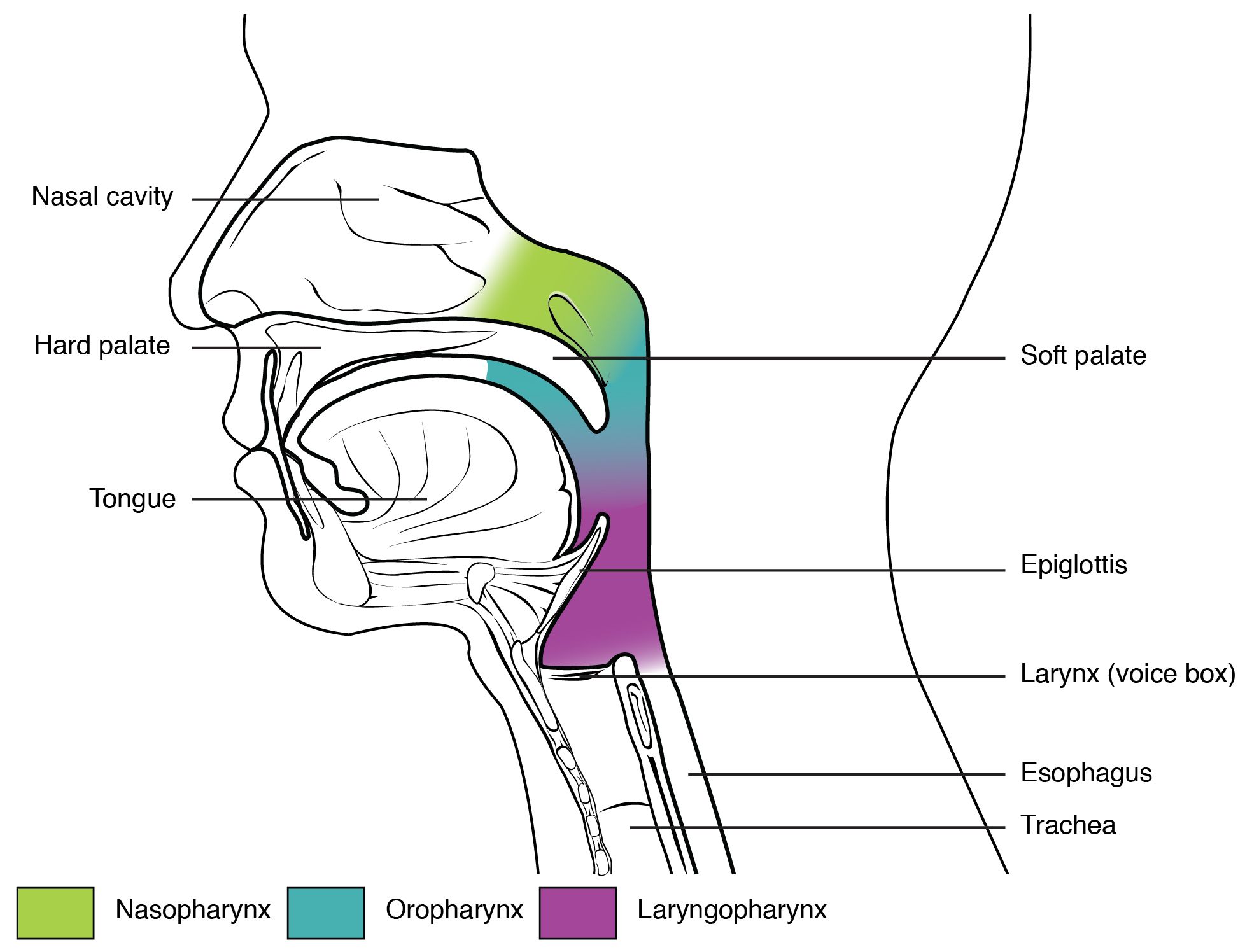
Parapharyngeal space
A deep neck space whose relations explain spread of infection and displacement of nearby structures.
Identify · Structures · Viva 2 references · 4 viva promptsRetropharyngeal space
Permits pharyngeal movement and is a potential route for infection.
Identify · Structures · Viva 2 references · 4 viva promptsDanger space of neck
A potential route for descending infection into the thorax.
Identify · Structures · Viva 2 references · 4 viva prompts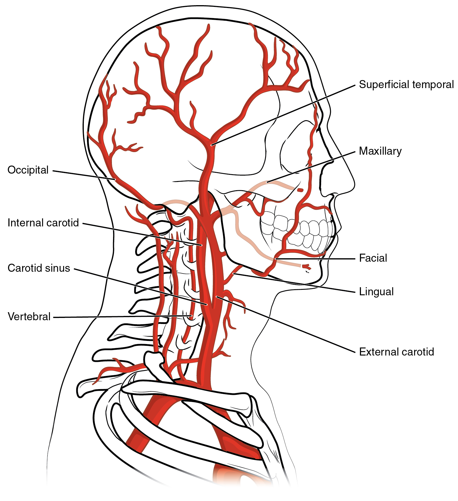
Carotid sheath
Organises the major neurovascular bundle of the neck.
Identify · Structures · Viva 3 references · 4 viva prompts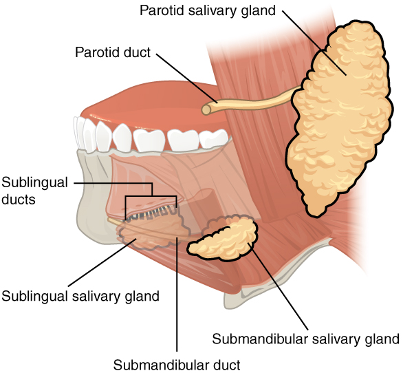
Sublingual space
Contains sublingual gland, submandibular duct and important neurovascular relations.
Identify · Structures · Viva 2 references · 4 viva promptsSubmandibular space
A potential site for spread of odontogenic infection.
Identify · Structures · Viva 2 references · 4 viva promptsSubmental space
A midline floor-of-mouth-related space.
Identify · Structures · Viva 2 references · 4 viva promptsParotid gland
Largest major salivary gland; predominantly serous secretion.
Identify · Structures · Viva 2 references · 4 viva promptsParotid duct (Stensen)
Conducts parotid secretion to the oral vestibule.
Identify · Structures · Viva 2 references · 4 viva promptsSubmandibular gland
Produces mixed, predominantly serous saliva.
Identify · Structures · Viva 2 references · 4 viva promptsSubmandibular duct (Wharton)
Carries submandibular saliva into the oral cavity.
Identify · Structures · Viva 2 references · 4 viva promptsSublingual gland
Produces mixed saliva, predominantly mucous.
Identify · Structures · Viva 2 references · 4 viva promptsMinor salivary glands
Maintain local mucosal lubrication through short ducts.
Identify · Structures · Viva 2 references · 4 viva promptsNasopharynx
Conducts air and communicates with the middle ear through auditory tubes.
Identify · Structures · Viva 2 references · 4 viva promptsOropharynx
A shared passage for swallowed material and air.
Identify · Structures · Viva 2 references · 4 viva promptsLaryngopharynx
Directs swallowed material around the laryngeal entrance toward the oesophagus.
Identify · Structures · Viva 2 references · 4 viva prompts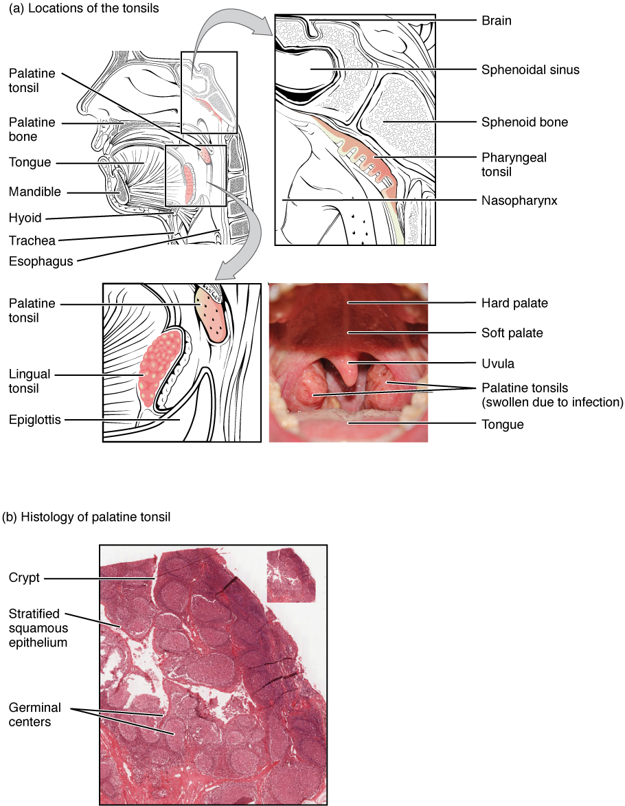
Palatine tonsil
Part of Waldeyer's lymphoid ring.
Identify · Structures · Viva 2 references · 4 viva prompts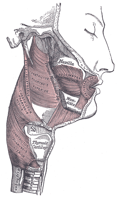
Pharyngeal constrictor muscles
Propel a swallowed bolus inferiorly.
Identify · Structures · Viva Focused view + full reference · 4 viva prompts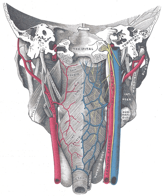
Stylopharyngeus muscle
Elevates and widens the pharynx during swallowing.
Identify · Structures · Viva Focused view + full reference · 4 viva promptsSoft palate
Separates nasopharynx from the swallowing pathway when elevated.
Identify · Structures · Viva 2 references · 4 viva prompts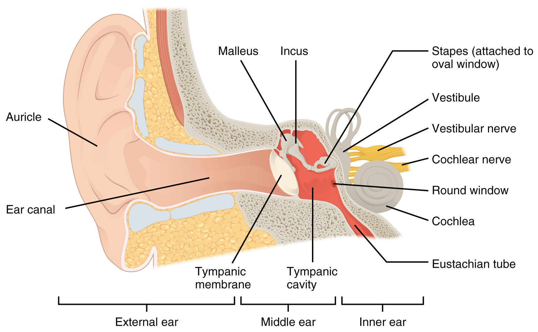
Pharyngotympanic (auditory) tube
Ventilates the middle ear and helps equalise pressure.
Identify · Structures · Viva 2 references · 4 viva prompts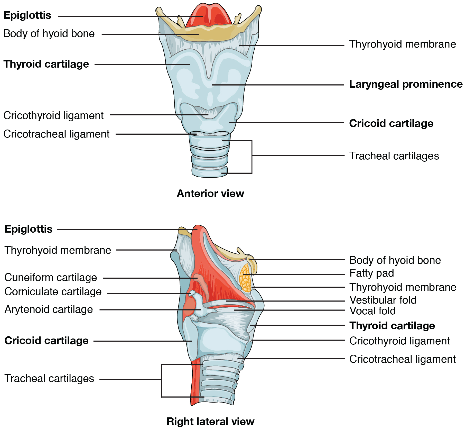
Epiglottis
Contributes to protection of the laryngeal inlet during swallowing.
Identify · Structures · Viva 2 references · 4 viva promptsCricoid cartilage
Supports the larynx and forms joints for laryngeal movement.
Identify · Structures · Viva 2 references · 4 viva promptsArytenoid cartilages
Position the vocal folds during breathing and phonation.
Identify · Structures · Viva 2 references · 4 viva prompts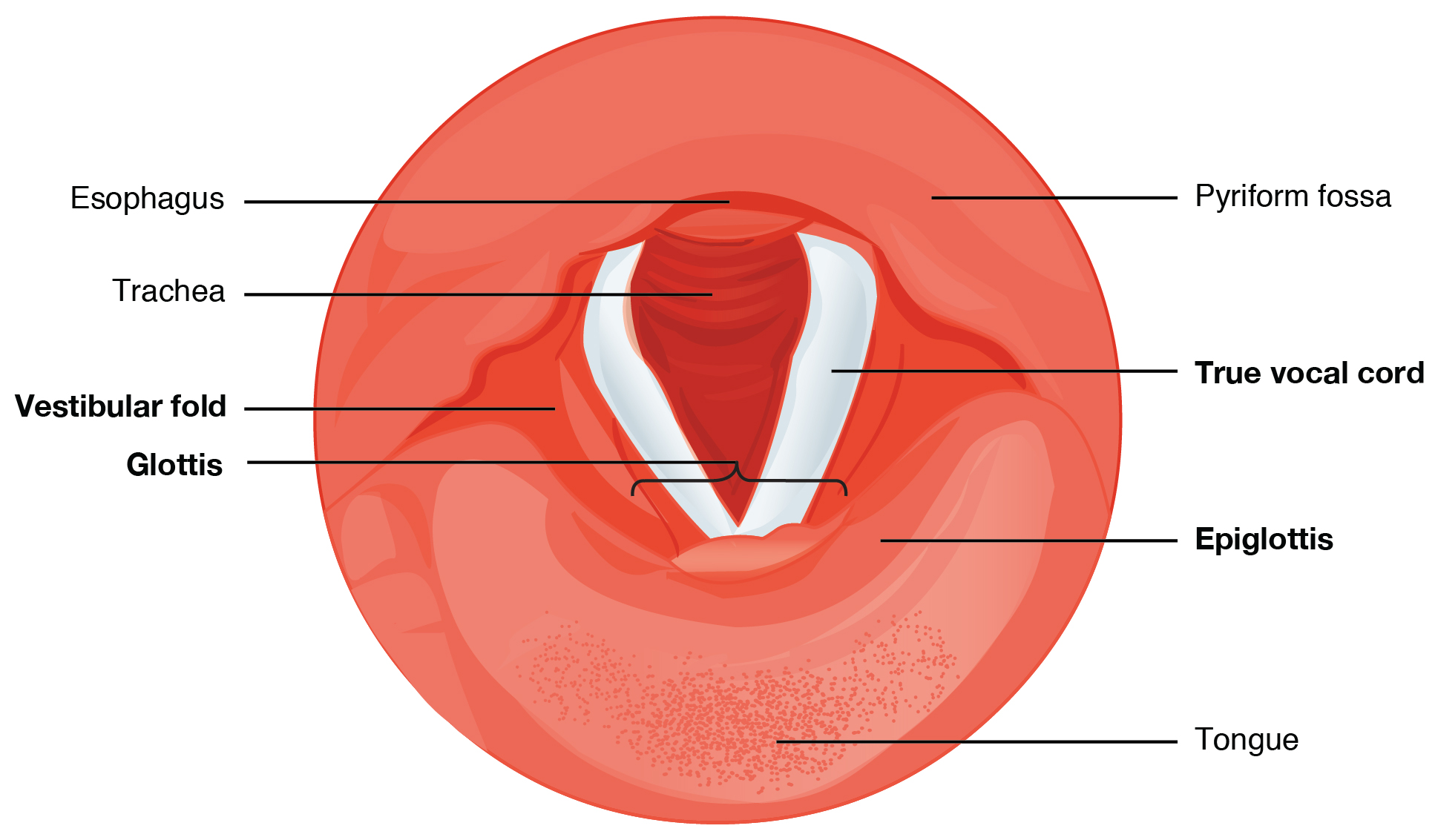
True vocal folds
Vibrate in phonation and participate in airway closure.
Identify · Structures · Viva 2 references · 4 viva promptsPosterior cricoarytenoid
Opens the rima glottidis by abducting the vocal folds.
Identify · Structures · Viva 2 references · 4 viva promptsCricothyroid muscle
Tenses and lengthens the vocal folds.
Identify · Structures · Viva 2 references · 4 viva promptsAuricle (pinna)
Collects sound into the external acoustic meatus.
Identify · Structures · Viva Focused view + full reference · 4 viva promptsTympanic membrane
Transfers acoustic vibration to the ossicular chain.
Identify · Structures · Viva Focused view + full reference · 4 viva promptsTympanic cavity
Conducts sound through the middle-ear ossicles.
Identify · Structures · Viva Focused view + full reference · 4 viva promptsMalleus
Receives vibration from the tympanic membrane.
Identify · Structures · Viva Focused view + full reference · 4 viva promptsIncus
Links malleus and stapes.
Identify · Structures · Viva Focused view + full reference · 4 viva promptsStapes
Transmits vibration into inner-ear fluid.
Identify · Structures · Viva Focused view + full reference · 4 viva promptsOval window
Receives mechanical energy transmitted by the ossicles.
Identify · Structures · Viva Focused view + full reference · 4 viva promptsRound window
Allows compensatory movement of cochlear fluid.
Identify · Structures · Viva Focused view + full reference · 4 viva promptsCochlea
Contains the sensory apparatus for hearing.
Identify · Structures · Viva Focused view + full reference · 4 viva promptsVestibule, utricle and saccule
Otolith organs detect linear acceleration and head orientation relative to gravity.
Identify · Structures · Viva Focused view + full reference · 4 viva promptsSemicircular canals
Detect angular acceleration through the membranous semicircular ducts.
Identify · Structures · Viva Focused view + full reference · 4 viva promptsStapedius
Modulates stapes movement.
Identify · Structures · Viva 1 reference · 4 viva promptsTensor tympani
Tenses the tympanic membrane through the malleus.
Identify · Structures · Viva 1 reference · 4 viva prompts
Olfactory nerve (I)
Special sensory smell; numerous filaments pass through the cribriform plate.
Identify · Structures · Viva Focused view + full reference · 4 viva promptsOptic nerve (II)
Carries vision and the afferent limb of the pupillary light reflex.
Identify · Structures · Viva Focused view + full reference · 4 viva promptsOculomotor nerve (III)
Supplies most extraocular muscles, levator palpebrae and preganglionic parasympathetic fibres to the ciliary ganglion.
Identify · Structures · Viva Focused view + full reference · 4 viva promptsTrochlear nerve (IV)
Motor supply to superior oblique.
Identify · Structures · Viva Focused view + full reference · 4 viva prompts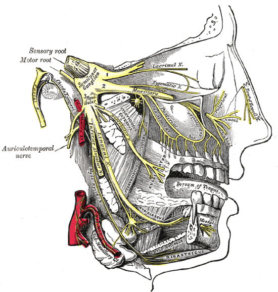
Ophthalmic division (V1)
Sensory division supplying forehead, cornea and related upper facial regions.
Identify · Structures · Viva Focused view + full reference · 4 viva promptsMaxillary division (V2)
Sensory division for midface, upper teeth, palate and nasal regions.
Identify · Structures · Viva Focused view + full reference · 4 viva promptsMandibular division (V3)
Mixed division supplying lower facial sensation and first-arch muscles.
Identify · Structures · Viva Focused view + full reference · 4 viva promptsAbducens nerve (VI)
Motor supply to lateral rectus.
Identify · Structures · Viva Focused view + full reference · 4 viva promptsFacial nerve (VII)
Mixed nerve for facial expression, taste and parasympathetic pathways.
Identify · Structures · Viva Focused view + full reference · 4 viva promptsVestibulocochlear nerve (VIII)
Special sensory nerve for hearing and equilibrium.
Identify · Structures · Viva Focused view + full reference · 4 viva promptsGlossopharyngeal nerve (IX)
Mixed nerve with pharyngeal sensation, stylopharyngeus motor and parotid parasympathetic functions.
Identify · Structures · Viva Focused view + full reference · 4 viva promptsVagus nerve (X)
Mixed nerve supplying the pharynx, larynx and thoracoabdominal viscera.
Identify · Structures · Viva Focused view + full reference · 4 viva promptsAccessory nerve (XI)
Spinal accessory fibres supply sternocleidomastoid and trapezius.
Identify · Structures · Viva Focused view + full reference · 4 viva promptsHypoglossal nerve (XII)
Motor nerve to intrinsic and most extrinsic tongue muscles.
Identify · Structures · Viva Focused view + full reference · 4 viva promptsLingual nerve
Carries general sensation from anterior two-thirds of tongue and receives chorda tympani fibres.
Identify · Structures · Viva 3 references · 4 viva promptsInferior alveolar nerve
Sensory supply to lower teeth, with a mylohyoid motor branch before canal entry.
Identify · Structures · Viva 3 references · 4 viva promptsAuriculotemporal nerve
Sensory nerve that also conveys postganglionic parasympathetic fibres to parotid.
Identify · Structures · Viva 3 references · 4 viva promptsChorda tympani
Carries taste from anterior tongue and preganglionic parasympathetic fibres for submandibular/sublingual glands.
Identify · Structures · Viva 3 references · 4 viva prompts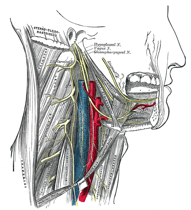
Superior laryngeal nerve
Divides into sensory internal and mainly motor external branches.
Identify · Structures · Viva 2 references · 4 viva promptsRecurrent laryngeal nerve
Supplies most intrinsic laryngeal muscles and sensation below vocal folds.
Identify · Structures · Viva 2 references · 4 viva promptsAnsa cervicalis
Cervical motor loop supplying sternohyoid, sternothyroid and omohyoid.
Identify · Structures · Viva 2 references · 4 viva promptsCervical plexus
Supplies neck skin and muscles and contributes to the phrenic nerve.
Identify · Structures · Viva 2 references · 4 viva promptsCervical sympathetic trunk
Sympathetic pathway for head and neck structures.
Identify · Structures · Viva 2 references · 4 viva promptsOtic ganglion
Parasympathetic relay for parotid gland.
Identify · Structures · Viva 3 references · 4 viva promptsPterygopalatine ganglion
Parasympathetic relay for lacrimal, nasal and palatal secretomotor pathways.
Identify · Structures · Viva 3 references · 4 viva promptsSubmandibular ganglion
Parasympathetic relay associated with the floor of mouth.
Identify · Structures · Viva 3 references · 4 viva promptsCommon carotid artery
Main arterial trunk that usually bifurcates near the upper border of thyroid cartilage.
Identify · Structures · Viva 3 references · 4 viva promptsExternal carotid artery
Supplies much of the external head and neck.
Identify · Structures · Viva 2 references · 4 viva prompts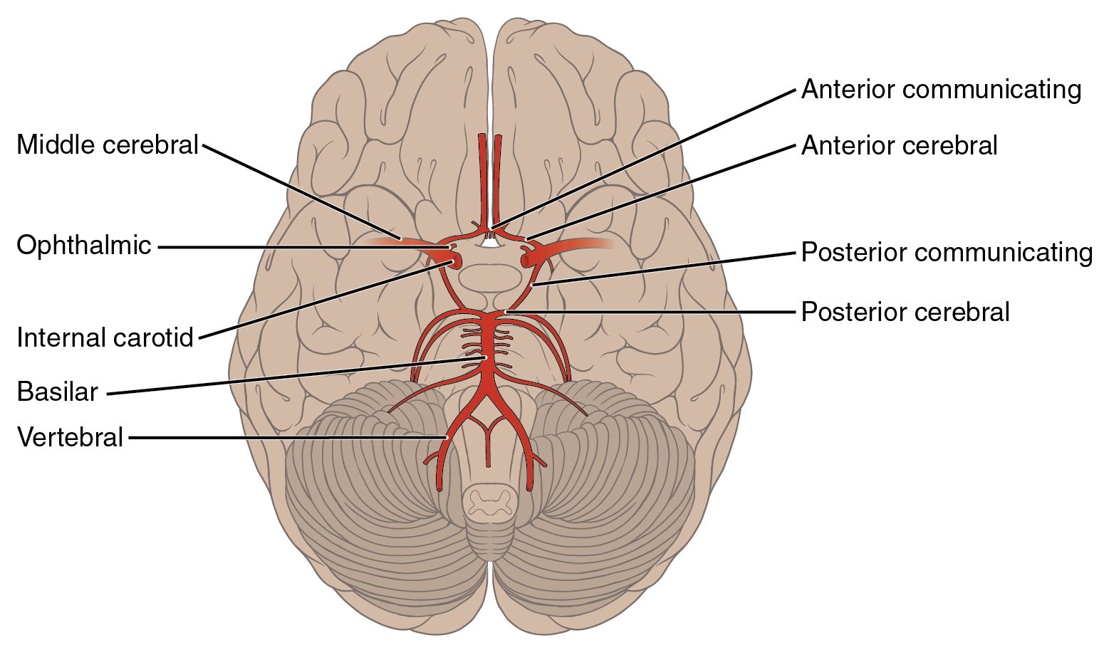
Internal carotid artery
Supplies intracranial structures and orbit through its branches.
Identify · Structures · Viva 2 references · 4 viva promptsCarotid sinus and body
Sinus is a baroreceptor region; carotid body is a peripheral chemoreceptor.
Identify · Structures · Viva 2 references · 4 viva promptsFacial artery
Tortuous artery supplying face and related pharyngeal/palatal structures.
Identify · Structures · Viva 2 references · 4 viva promptsLingual artery
Major artery of tongue and floor-of-mouth regions.
Identify · Structures · Viva 2 references · 4 viva promptsMaxillary artery
Terminal ECA branch supplying deep face, teeth, dura and nasal/palatal regions.
Identify · Structures · Viva 2 references · 4 viva promptsMiddle meningeal artery
Major dural arterial supply.
Identify · Structures · Viva 2 references · 4 viva promptsSuperficial temporal artery
Supplies temporal scalp and adjacent regions.
Identify · Structures · Viva 2 references · 4 viva promptsVertebral artery
Contributes to posterior cerebral circulation.
Identify · Structures · Viva 2 references · 4 viva prompts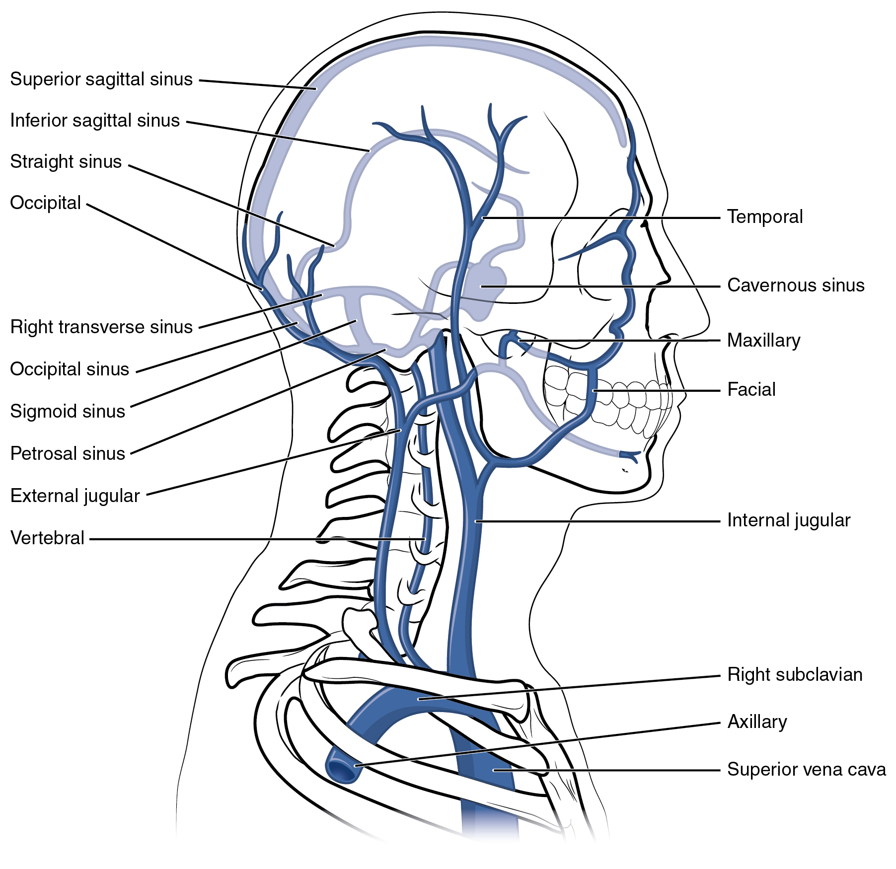
Internal jugular vein
Major venous drainage pathway from brain, deep face and neck.
Identify · Structures · Viva 3 references · 4 viva promptsExternal jugular vein
Drains superficial head and neck territories.
Identify · Structures · Viva 2 references · 4 viva promptsRetromandibular vein
Connects deep facial and superficial venous drainage.
Identify · Structures · Viva 2 references · 4 viva promptsPterygoid venous plexus
Venous network of the infratemporal region.
Identify · Structures · Viva 2 references · 4 viva prompts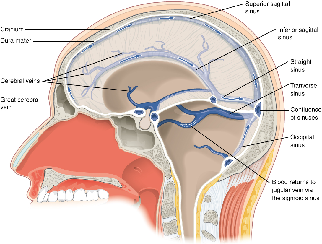
Cavernous sinus
Paired dural venous space with important neurovascular relations.
Identify · Structures · Viva 2 references · 4 viva prompts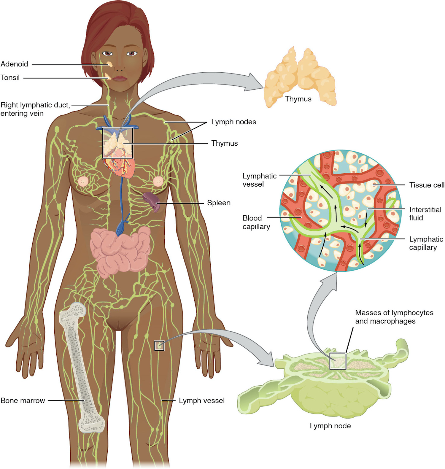
Deep cervical lymph nodes
Receive drainage from many head-and-neck lymphatic groups.
Identify · Structures · Viva 2 references · 4 viva prompts